No Research experience? Don’t live on campus? Want your own hours?
This project allows students to gain research experience at home and on their own time.
No experience needed!
Project Details:
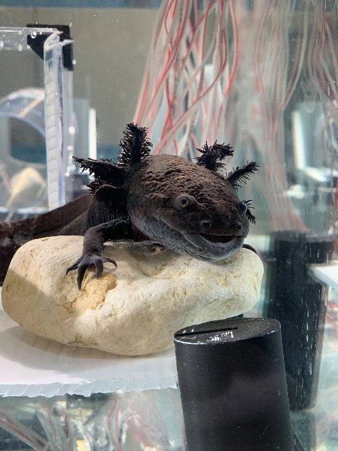
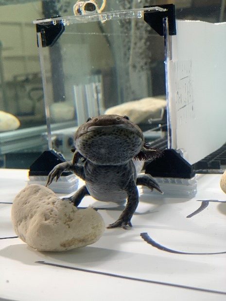
In the Astley lab, our primary focus is in the field of biomechanics, where we integrate animal biomechanics and morphological features. This project looks to expose the axolotl single leg kinematics when they are walking underwater. Tiered mentoring students will be digitizing videos of axolotls walking. Students will be exposed to scientific research and to lab settings. Dependent on mentee’s schedule, student can attend laboratory meetings, which consist of Astley lab members reviewing published scientific papers.
Click here for other information about Dr. Astley’s lab.
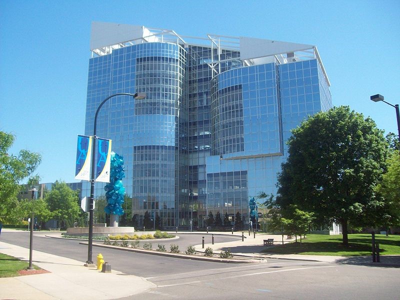
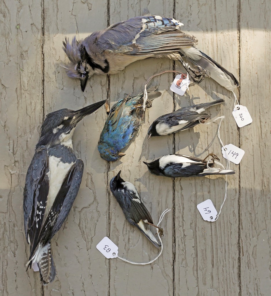
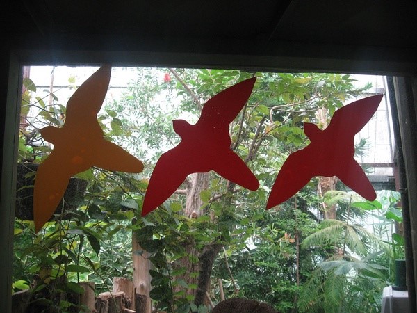
Good year polymer center, the building where our lab is located, has had disproportionally high bird strike incidents compared to any other building on campus or even the greater Akron area (unpublished data from the Akron Zoo). This is due to the predominantly glass design of the build that make it invisible to birds. We are hoping to mitigate that fact using low cost methods such artful decals decorations that add to the appeal of the building while protecting the birds. The undergraduate researcher for this project will be responsible for finding cost effective materials, designing the decals and lastly helping to put together a proposal to execute the plan. This is a continuation of a project that has been monitoring the number of dead birds around our building for the last couple of years.
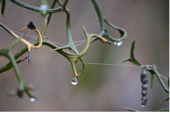
Vibrations are incredibly useful to spiders—it’s through vibrations that spiders are able to detect and locate prey, communicate with each other, and perceive their immediate surroundings. Many spiders rely on the webs they build to transmit this information, and the properties of those webs determine how quickly and efficiently they can detect and respond to different signals.
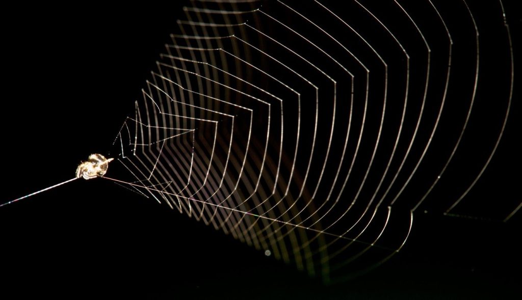
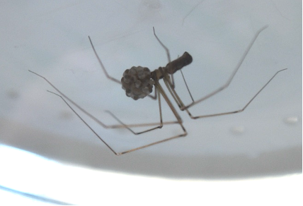
The slingshot spider (genus Theridiosoma) builds an orb-shaped web which it tightens from the center, transforming it into a conical, energy-loaded snare. When it detects flying prey, the spider launches the structure forward, intercepting any insects in its path.
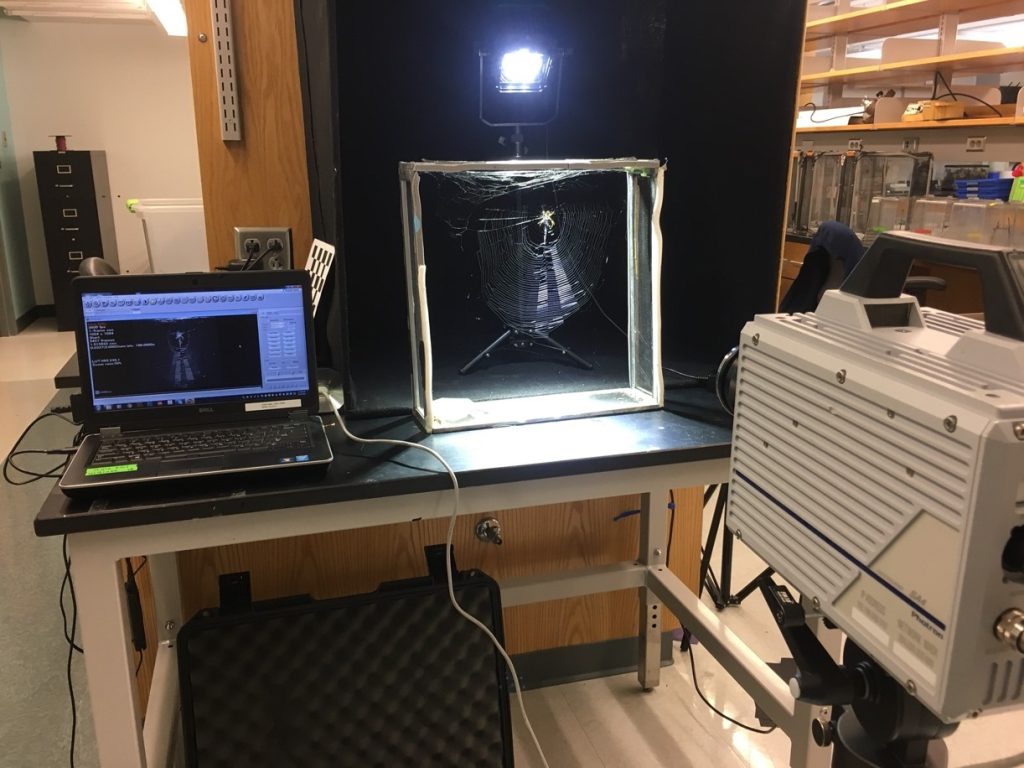
The unique characteristics of this system—the web shape, the active prey capture mechanism, and the spider’s ability to detect prey before the web touches it—make it ideal for studying vibration transmission. In this project, I aim to determine 1) how the three-dimensional shape of Theridiosoma webs may aid in transmitting vibrations and 2) the mechanism through which the spider detects its prey (either through airborne or web-borne vibrations).
In this project, you’ll gain experience with…
Field- and lab-based research
Invertebrate collection and care
High-speed videography and vibration-monitoring techniques
Mechanical testing and material science
Spider biology and behavior
Physics of vibration
Statistical analysis and data interpretation
Presentation in a professional setting and/or publication
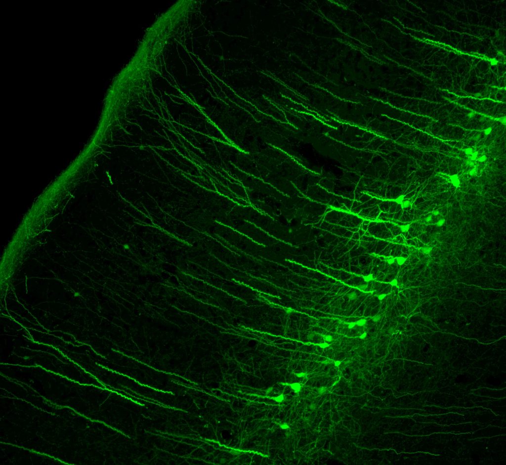
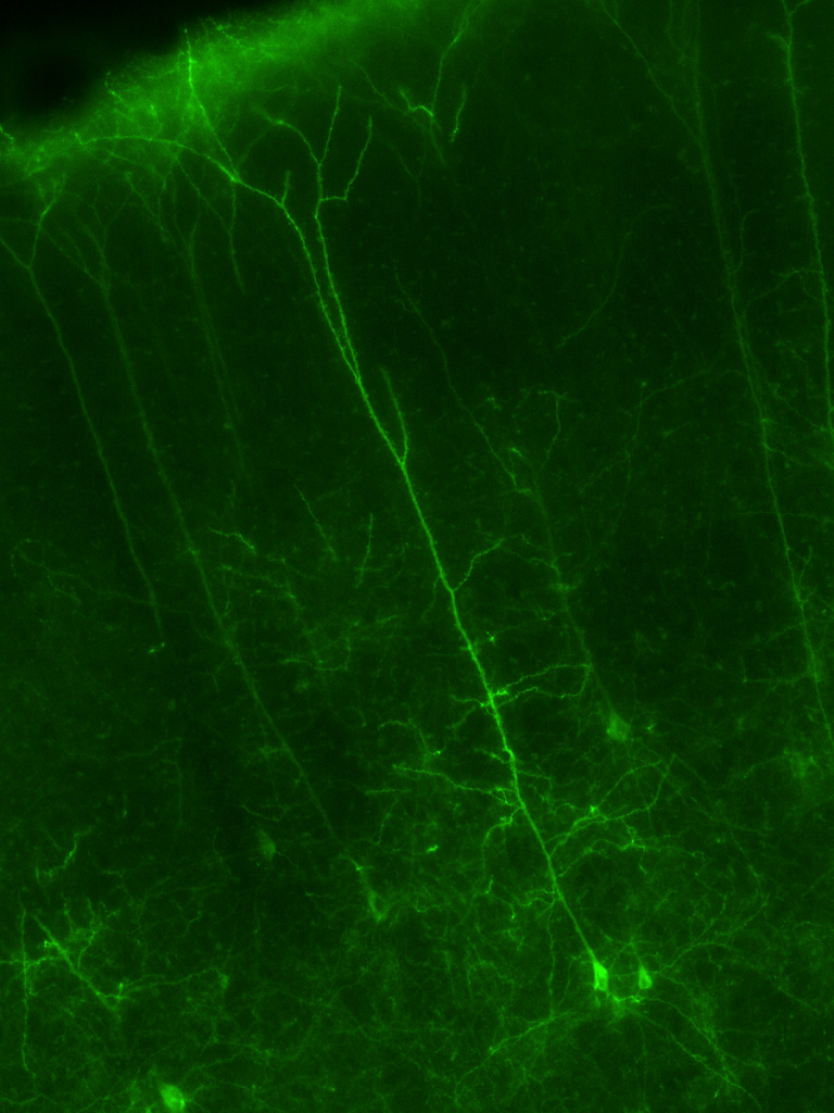
The Schofield Lab uses microscopy to study pathways in the brain, especially pathways important in hearing and attention that use acetylcholine as a neurotransmitter. We inject fluorescent tract tracers into auditory brain areas in mice and guinea pigs, then study pathways using a fluorescent microscope. We also fluorescently label brain cells with immuno-staining, a technique where antibodies are used to recognize proteins in the brain. These techniques allow us to map brain circuits that might explain why hearing your name in a crowded room grabs your attention, or why you can easily sleep through your partner’s snoring but awaken immediately at an unfamiliar noise. The photo below shows cells in the auditory cortex that make descending projections to the brainstem.GP799 1:7-1 left AC AAVrg in L IC; scale 250 um
Benefits to students:
Observe or participate in tracer injection surgeries
Participate in cutting of brains on a microtome and immuno-staining of brain sections
Learn how to use a fluorescence microscope and fluorescence microscopy software
Opportunity to participate in weekly Schofield Lab meetings
Access to NEOMED Hearing Research Group seminars and journal clubs
Skills needed:
None! We are happy to teach you everything you need to know to work in The Schofield Lab with us. All you need is an eagerness to learn and participate.
To read more about research in the Schofield lab, visit our website at www.schofieldlab.org.
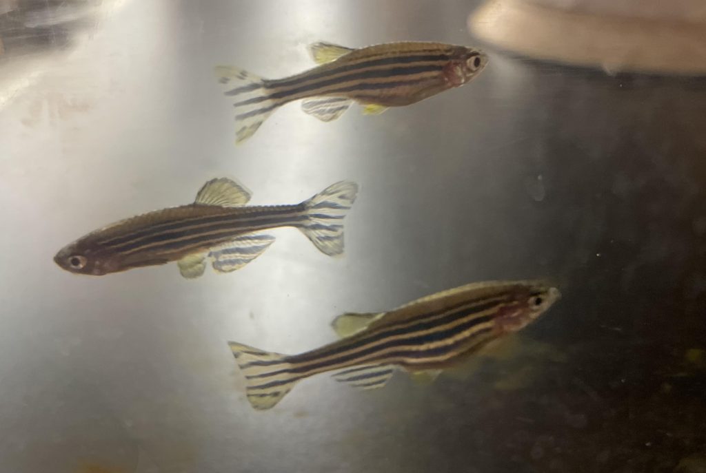
We are working towards understanding how different levels of macronutrients may impact the physiology of zebrafish. More specifically, we want to first see if when presented with foods that have different levels of major macronutrients (carbohydrate, fats, protein), will the fish choose one over the others or show any type of preference. From there, we want to specifically feed some fish one diet while others receive the alternative options and then run tests to understand how those feeding choices impact their behavior, how many eggs females will produce, and potentially the developmental rate of offspring and their cardiovascular development. Zebrafish are a great model organism for this type of research because of the ease of raising them, high fecundity, and the ability to observe the early development of the cardiovascular system, to name a few. Projects are continuously evolving, and potential students would gain valuable experience in animal care, as well as the importance of trouble shooting in science research.
Click here for more information on Dr. Bagatto’s lab.
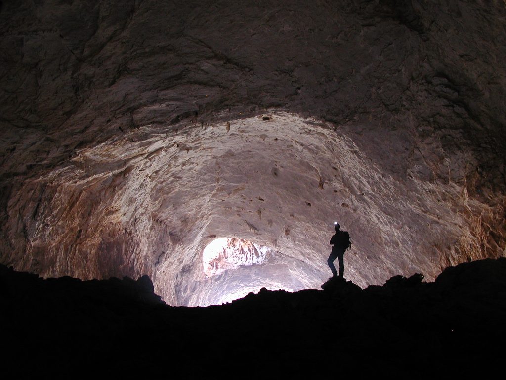
Background: Understanding the physical and chemical limits of life on Earth is an essential first step to understanding the possibility of extraplanetary life (astrobiology). Life requires energy, and the primary sources are chemical and light energy. Phototrophs harvest light energy between 400-700 nm wavelengths while a few species, such as purple bacteria, can harvest light energy from the lower energy near-infrared region of the spectrum (700-973 nm) using different photosynthetic and photoreactive proteins. Additional research into life that can survive in various light conditions (energy from even lower wavelengths and low intensities) is required to understand the lowest limits of light needed to support life.
OLYMPUS DIGITAL CAMERA
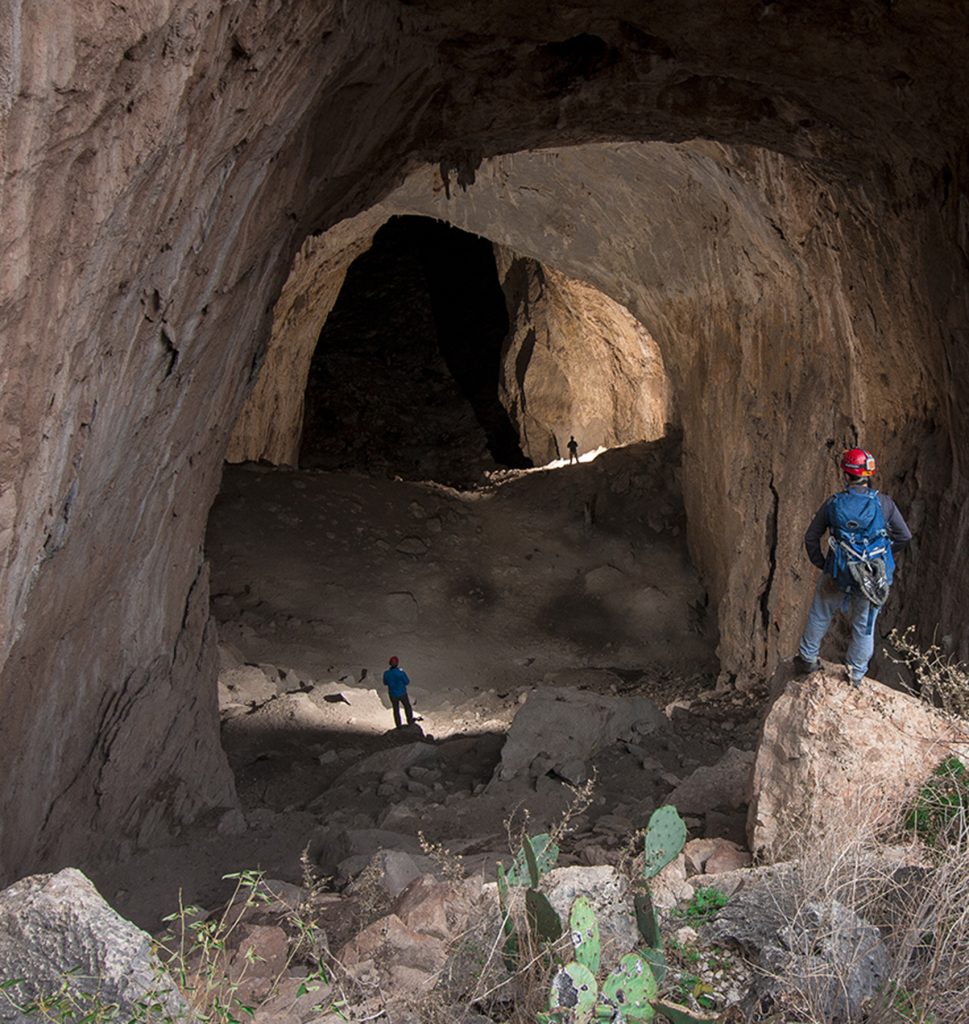
Terrestrial caves can be considered potential sites to carry out such research as they naturally demonstrate a range of light energy gradients (both light wavelengths and the amount of light energy per unit area) in their entrances. This leads to the stratification of different microbial communities that are adapted to use the available light sources depending on where they live. Surprisingly, even in the darkest zones of caves, longer wavelengths are available due to the reflection and refraction of light bouncing deeper into the caves. In 2019, we identified cyanobacteria even in the dark zones of caves in New Mexico that can harvest these lower energy near-infrared regions for photosynthesis under extremely low light levels. Thus, we can observe light-harvesting activities of life in caves in the complete dark where a light source is required to see your hand in front of your face.
Our Project: Our project aims to understand the limits of intensity and wavelength of light that can support life, using cave entrances as a novel study site. Samples will be analyzed for microbial community composition of known phototrophic species using 16S rRNA. The presence of genes involved in photosynthetic pigments and photo-reactive protein production will be identified using metagenomics. The expression of pigments will be analyzed using different analytical methods and instruments, including the pigment extractions using organic solvent partitioning and HPLC, ART-FTRI, GC-MS, Raman spectroscopy, and fluorescent absorption and emission data. Culture-based techniques under cave-relevant light conditions will be used to determine whether phototrophy is supported under the conditions found in the cave. Finally, direct cell counts will be carried out in each sample to estimate the relative abundance of available light energy-supported life under each light condition until we determine the limits of light-supported growth. Together these results should help us estimate the initial light energy limits necessary to support life in this model system.
What you can Learn (but not limited to): Microbiology techniques and culture methods, making different types of media, sample preparation for different analytical methods, experimental designs, and the use of various laboratory instruments such as pH meter, autoclave, pipettes, centrifuge, and microscopy.
Click here for more information about Dr. Barton’s lab
Research Area
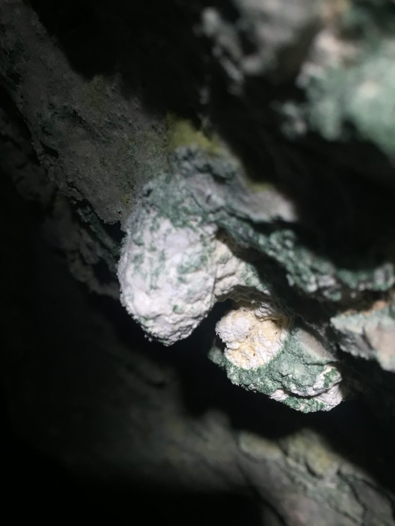
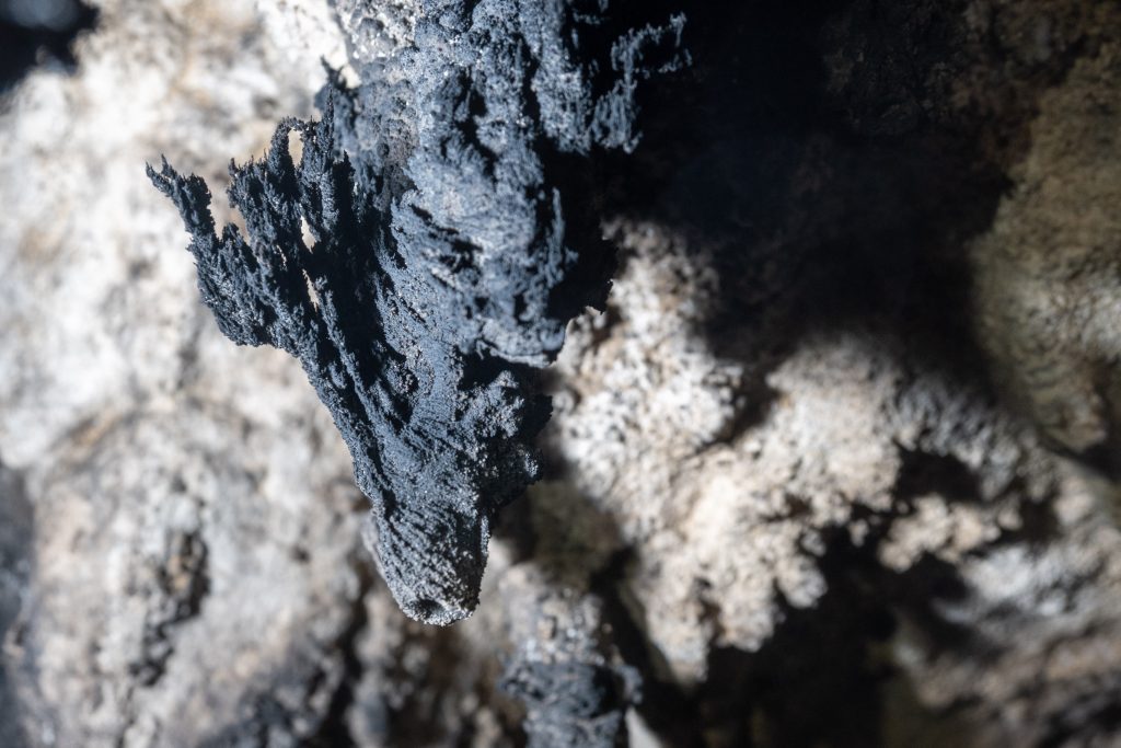
We study the interplay between the organic (microbes) and the inorganic (minerals) at the microscopic scale. Through a combination of metabolic and geochemical pathways, bacteria can initiate and influence mineral growth and/or decay. These processes are often influenced by cave climatology, creating extremely rare types of speleothems (calcium carbonate structures). Caves host a diverse range of microbial communities in close association with these speleothems, making these environments important sources of scientific discovery relating to mineralogy and geomicrobiology. We aim to investigate the mineralogy unusual speleothems collected from a remote cave and search for evidence of microbial influences in its formation.
Current Project
I am seeking a student interested in helping me investigate the mineralogical and microbiological aspects of cave-derived specimens using a combination of microscopy and geochemical techniques. Student will utilize a range of analysis techniques potentially including X-ray powder diffraction, Raman spectroscopy, and microscopy. Students will begin to gain a practical understanding of mineralogical research, develop a range of laboratory skills, and assist in planning and carrying out experiments.
Fig. 1: carbonate speleothem showing strong climatological influence and potentially microbial influence. Photo by Dr. Jean Krejca, Zara Environmental, LLCClick here for more information about Dr. Barton’s lab
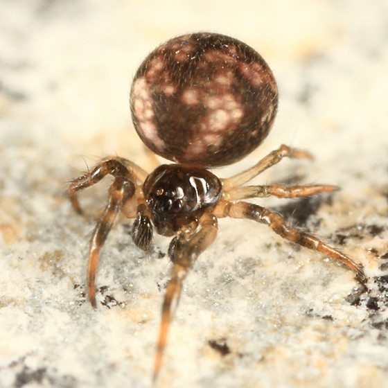
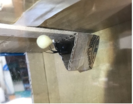
Spiders’ egg sacs are woven from multiple types of spider silk and offer protection to developing embryos and spiderlings. There is extreme diversity among egg sacs across species in shape, color, texture, and habitat, and little is currently known about how these variations affect spider development.
Extreme water loss is one threat to developing spiders that can cause them to desiccate, or dry out. I am studying whether spider egg sac materials protect against desiccation of developing spiders by preventing water from escaping the inside of the egg sac. I would also like to explore which characteristics of the egg sac silk formations contribute to lower or higher transmission rates through the material.
Skills you will acquire:
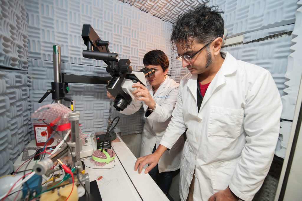
Jeffrey Wenstrup, Ph.D., Sharad Shanbhag, Ph.D., and Mahtab Tehrani, Ph.D.
Our Work
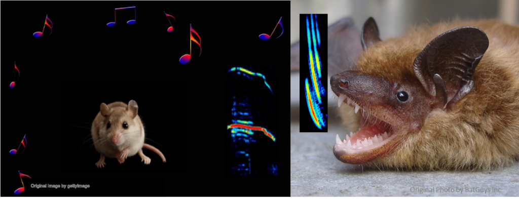
Vocalizations reflect our emotions. As we listen to another’s vocal signals, we respond by assigning them a meaning determined by the vocalization itself, our previous experience, and our own emotional state. This in turn affects the way that we respond to a person’s vocal signals—by our posture, facial gestures, movement, and speech. Our goal is to understand how the brain shapes our responses to these vocal signals.
Our approachBrain regions of interest:
Our focus is on the amygdala, a brain center that integrates sensory inputs with information about our previous experiences and our internal state. It then assesses the meaning of new sensory information and “decides” on the appropriate behavioral responses. We also study the auditory centers that feed information to the amygdala about social vocalizations.
Our models
We use bats and mice as models to study these processes. Bats are sound experts that use vocalizations to both communicate and to catch prey and navigate through echolocation. Mice are acoustic generalists that integrate acoustic and other sensory information during social interactions. Both models provide valuable insights into the mechanisms underlying acoustic communication and emotions.
Our techniques:
We record and analyze the social vocalizations of bats and mice to understand how they communicate their emotional state through social vocalizations. These analyses help us to design vocal stimuli to study the animals’ behavioral and brain responses to emotion-containing vocalizations.
Our neural studies use a combination of anatomical tracing, optogenetics, imaging, electrophysiology, microdialysis, and pharmaceutical techniques. These help us to describe how amygdala neurons respond to vocalizations, which brain areas drive the amygdalar responses to vocalizations, and how modulatory neurochemicals shape these neuronal responses and behavioral reactions.
Research Projects involving undergraduates
• Describing vocal behavior in mice
• Analyzing circuitry related to emotions and vocalizations
• Analyzing neural responses to vocalizations
Publicationshttps://scholar.google.com/citations?hl=en&user=VxcRjv0AAAAJ&view_op=list_works&sortby=pubdateClick here for more information on the Wenstrup lab.
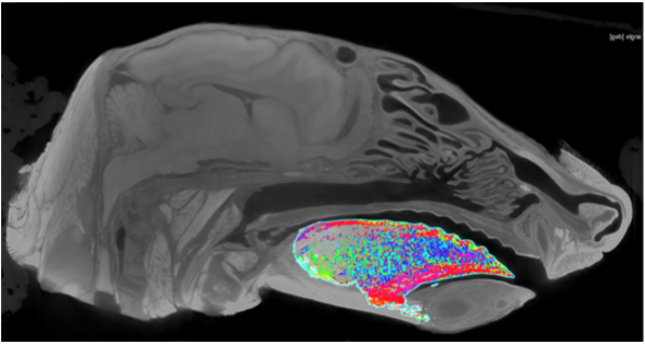
The Olson lab is interested in the form and function of the mammalian feeding system. We look at this through studies of mammalian diversity and evolution, as well as using animal models to better understand human disorders. A multitude of anatomical structures are involved in the feeding apparatus, including the tongue. These muscles have complex orientations that are hard to dissect and measure. Therefore, we use diceCT scanning methods, where the soft-tissues are differentially stained with an iodine-based contrast enhancing stain, to digitally dissect these structures. Digital dissection preserves the 3D orientation of structures while allowing us to visualize and quantify the structures of interest.
Primary Goals
Digitally dissect the muscles involved in chewing (multiple individuals and species have been scanned).
Determine the fiber orientations of the tongue.
Potential to develop an analysis method to compare different species.
Potential to apply anatomical data of the tongue to in vivo biomechanics of the tongue.
Skills (no experience necessary):
Anatomical imaging – learn about and look at CT scans and x-ray imaging. There may be opportunities to participate in collecting this data.
Digital dissection methods using image processing software like Slicermorph and VGstudio.
Interact with and learn anatomy and biomechanics in an applied context.
Contrast-enhanced staining methods in the wet lab.
Possibility of traditional gross dissections.
Data visualization and statistical analysis in R.
Participate in lab meetings and scientific discussions.
Participate in the Biological Undergraduate Research Symposium.