The effects of invasive and naturalized worms on plant growth

[Past Projects]

Dr. Randall Mitchell and Cynthia Yoder


The Mitchell lab studies various ecological questions, from plants to animals and the human interactions with the environment. For this project, we will be studying the way worms change soil structure and how they may be affecting plants within the soils in which they exist.
In this project we will be conducting a laboratory experiment using three species of worms and produced castings to grow plants, measuring the changes in growth during this process, as well as changes in flower production and others. You will learn about the use of composting worms in agriculture and home gardening, hands-on scientific study of collecting and analyzing data, soil and worm sampling methodology, sample preparation and preservation, and applied scientific work, along with soil processes and more.
Click here for more information about the Mitchell lab.

Zebrafish as a model for bone remodeling

[Past Projects]

Dr. Richard Londraville and Kathryn Bisaha


Castranova, D., Weinstein, B., Samasa, B., n.d. Dorsal view of bones and scales (blue) and lymphatic vessels (orange) in a juvenile zebrafish | 2020 Photomicrography Competition. 
The Londraville lab seeks to explore the relationship between endospanin, leptin and bone in zebrafish through the comparison of various mutant strains. In this project students will explore how some variable (e.g., age, weight, mutation) affects bone formation and bone resorption in zebrafish. You will gain hands-on experience in the following areas: zebrafish handling and care, ImageJ analysis, microscopy, microinjection, and protein assays.
Click here for more information on Dr. Londraville’s lab.

Digital tracing neurons in a mouse model of glaucoma

[Past Projects]

Dr. Jordan Renna and Sara Chaszeyka


Our lab is studying the effects of the debilitating eye disease glaucoma on melanopsin ganglion cell neurons in the mouse retina. Melanopsin ganglion cells send light-evoked signals to the brain to regulate our circadian rhythms. Glaucoma is a medical condition in which eye pressure increases to the point at which neurons connecting the eye to the brain begin to die.   We are looking for a brave soul to digitally trace fluorescently labeled melanopsin ganglion cell neurons.  These neurons are from retinas in various stages of glaucoma. Our hope is to one day resolve glaucoma and cure the leading cause of rodent blindness. Our lab motto: NO BLIND MICE!
Click here for more information on Dr.Renna’s lab.

Synaptic Connections between light detecting cells and interneurons in the zebrafish retina

[Past Project]

Dr. Jordan Renna and Matthew Tarchick


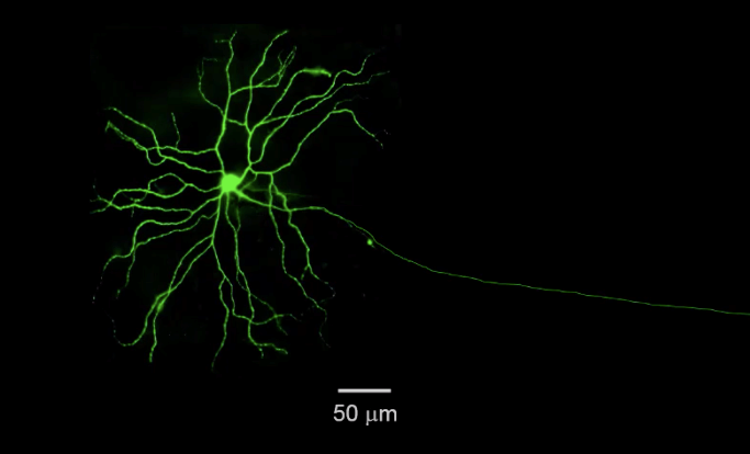
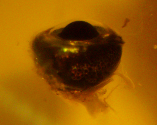
Our lab utilizes a procedure called Electroretinogram (ERG) to study the behavioral and physiological properties of the neural retina. This protocol has medical relevance and is routinely done by optometrists. It demonstrates the whole function of the retina and the function of several important cell populations. The A wave of the ERG is related to total output of light detecting cells (rods and cones). The B-wave of the ERG is related to the total output of the interneurons (or bipolar cells). These cells form a complex synapse that is needed for the transmission of visual information.  

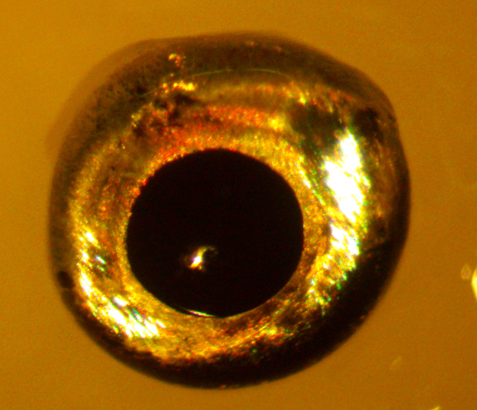
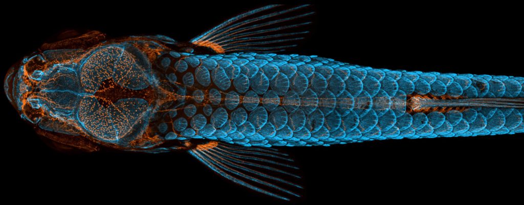
Our new project aims to classify how these connections are established and occur in zebrafish. Very few experiments have been done to measure the responsiveness of the zebrafish retina, so this approach is somewhat novel. The ease at which zebrafish can be genetically altered also opens more possible avenues for studying disease models and makes zebrafish a good model organism.  

We are looking for students to help run ERG experiments and conduct the analysis of those experiments.  


Click here for more information on Dr.Renna’s lab.

Biomechanics of Locomotion in an Animal Model of Preterm Infant Motor Development

[Past Projects]

Dr. Jesse Young (Department of Anatomy and Neurobiology, Northeast Ohio Medical University)


Preterm infants constitute 11% of all infants born in the United States.  Most of these infants show significant delays in motor development. However, the exact physiological mechanisms that account for these delays remain a mystery. Previous studies have hypothesized causes as disparate as early postnatal respiratory distress, poor cerebellar development, low muscle mass, and limits on muscle power production. In part, this gap in our knowledge stems from the integrative nature of locomotion itself.  Safe, efficient walking requires the coordinated output of multiple organ systems (respiratory, circulatory, nervous, musculoskeletal) to modulate the energetic and biomechanical demands of supporting and accelerating body mass. Only a longitudinal, multimodal study could provide the type of integrative physiological and mechanical data required to address the etiology of preterm infant motor delays. Such a study is not feasible in a compromised population like preterm human infants. Our lab has been developing the infant pig as an animal model of preterm human infant motor development. You would contribute to this goal by analyzing and interpreting previously collected data on walking, running, and standing in preterm and term infant pigs, contributing to our understanding of the underlying causes of preterm motor dysfunction. Skills you will develop:
  • Familiarity with modern biomechanical techniques used to analyze motor function and coordination in animals and humans
  • Use of MATLAB and R for data analysis and statistical interpretation
  • Opportunities to participate in weekly Biomechanics Journal Club meetings in the Department of Anatomy and Neurobiology at NEOMED and attend associated seminars

More about locomotor research in the Young Lab: www.younglaboratory.org.

‘I’m too freaked out to hear you!’ How early-life stress affects auditory perception and the brain

[Past Projects]

Merri Rosen PhD, Kate Hardy MS, and Matthew Sunthimer BS


It’s well-known that hearing problems in kids may cause later trouble with understanding complex sounds, such as rapid speech in a place with a lot of background noise. But our lab is the first to show that stress during development can cause similar problems. We’re funded by the National Institutes of Health to study the neural changes that can cause these stress-induced deficits in auditory perception. Many labs have studied the effects of early life stress (ELS) on the development of brain regions responsible for attention, learning, and related psychopathologies. However, nobody has examined whether auditory information is being encoded properly before it even reaches these higher-level brain regions. This is important, because children who grow up in low-socioeconomic, high stress environments are at risk for later problems with speech perception. Discovering what is going wrong mechanistically will help prevent and remediate these problems.
Our lab has shown that ELS affects auditory perception and neural activity in brain regions that encode sound. It turns out that ELS may be a particular problem when kids have ear infections that cause intermittent hearing loss – our data show that early hearing loss and stress together are much worse than either one alone! We use an animal model (the Mongolian gerbil) to study how early-life stress and hearing loss affect the perception of rapid changes in sound, and the underlying neural mechanisms. The Mongolian gerbil is a well-established auditory model, because gerbils hear well at frequencies that humans use, unlike mice or rats. Several projects are available in our lab: Seeing stress in the brain!
  • Background: Auditory perceptual problems from ELS and hearing loss may arise from changes in certain molecules that determine how neural circuits are wired during development. Measuring these elements will clarify whether deficits from ELS and hearing loss arise through similar or disparate mechanisms.
  • Objectives: To investigate changes in specific molecular markers within the auditory pathway across development. These changes are indicative of alterations in functionality and plasticity across development.
  • Methods: The effects of early stress and hearing loss on the brain will be identified using immunohistochemistry techniques which tag specific neuronal proteins with fluorescent markers. These markers can be visualized and quantified under a fluorescent microscope.
What stresses out our animals? Perception in action: Testing the limits of hearing
  • Background: Historically, mice and rats have been primarily used in stress research. To study stress in an established animal model of hearing, we are developing a new model of stress in the Mongolian gerbil.
  • Objective: To characterize the effects of the limited bedding model (LBM) on emotion, cognition, and auditory perception.
  • Methods: Implementation of the LBM model uses video recordings of breeder cages in a bedding-scarce environment. A combination of behavior tracking software and manual hand scoring of recordings will be used to characterize the new model. Various biomarkers will be measured to quantify stress markers, such as blood corticosterone and body weight. Effects on anxiety and cognition will be measured by standardized behavioral tests. Deficits in auditory perception will be measured using operant conditioning to train animals to respond when they discriminate between behaviorally-relevant sounds.
Livestreaming real-time brain activity: The Miniscope
  • Background: Our lab is working to set up an optical Miniscope, which allows us to visualize neuronal activity across a population of cells in real time, in freely-moving behaving animals.
  • Objectives: To contribute to getting this leading-edge technology set up in the lab.
  • Methods: Various skills will be learned while working closely alongside a lab member.
Benefits to student:
  • Receive training in experimental techniques used widely across research fields in biology and neuroscience
  • Learn how to apply critical thinking to big problems in neuroscience
  • Opportunity to join our lab as a PhD student or technician

Learn more about the Rosen lab.

Understanding the Microbial Activity involved in Forming Iron Caves in Brazil

[Past Projects]

Dr. John Senko and Melissa Mulford


Research Area: In the Carajas region of Brazil, banded iron formation (BIF) and its cap rock of canga are highly resistant to erosion and are poorly soluble. Despite this, over 3,000 caves have formed at their interface (one of these caves is shown in the photo). Our previous research has shown that microbial communities from these caves include bacterial species capable of Fe(III) reduction and the community is capable of extensive Fe(III) reduction. We hypothesize that microbial Fe(III) reduction transforms the insoluble Fe(III)-oxides into soluble Fe(II), which is then mobilized out the system, allowing caves and voids to form. We believe the Fe(II) may be stabilized in solution by a chemical interaction with silicates from the BIF. We are currently studying the process of Fe(II) and Si mobilization through microbially driven iron redox reactions and the effects dissolved silica has on iron redox reactions. We use bacterial cultures grown in both anaerobic and aerobic conditions with iron and silica. We then analyze these cultures with geochemical and biological analyses. You will learn many skills including but not limited to: microbial culturing and analyzing techniques, geochemical analysis methods, data processing methods, and experimental design.
Click here to learn more about Dr. Senkos’ lab.

Lipid modulation of membrane protein properties

[Past Projects]

Dr. Adam Smith and Grant Gilmore


Human cells produce hundreds of different lipid species, and the lipid complements of even closely related cells are diverse. Membrane lipid compositions are also highly susceptible to external inputs, especially cholesterol, polyunsaturated fatty acids (PUFAs), and gangliosides. In animal models, deprivation of ω-3 PUFAs leads to neurodevelopmental deficits in the offspring. However, the biomolecular connection between lipid composition and cellular function is not well-understood. In this project the student will conduct experiments to determine how lipid content is coupled to membrane protein function. Cultured cells will be supplemented with different lipid inputs, and the effects on membrane proteins will be measured using quantitative biophysical methods. This project is part of a collaborative research grant focused on neurological pathologies associated with brain lipid composition.
Click here to find out more about Dr. Smith’s research.

Silk properties of spider egg sacs

[Past Projects]

Dr. Todd Blackledge and Kate Karkosiak


Spiders’ egg sacs are made up of multiple types of spider silk and offer protection to developing embryos and spiderlings. Egg sacs exhibit extreme diversity across species in shape, color, texture, and placement, and little is currently known about the importance of these variations for appropriate spider development. I am interested in studying the effect of egg sac variations on the insulative properties, permeability, and other protective mechanisms of egg sac silk barriers related to their environmental or predatory threats.
Skills you will acquire: Knowledge about spider biology Knowledge about spider silk Field experience Research experience Scientific reading/writing
Click here for more information about Dr. Blackledge’s research.

Lights, Camera, Axolotl: A Behavioral Study

[Past Projects]

Dr. Henry Astley and Kaelyn Gamel


In the Astley lab, our primary focus is in the field of biomechanics, where we integrate animal biomechanics and morphological features. Specifically, this project transpires to study salamander behavior and how the axolotl interacts with its environment during underwater walking.  The neotenic salamander displays variable locomotive behaviors while interacting with the varying bottom substrate. Tests will be attained on different experimental substrates such as rocks, sticks, sand, other debris. Dependent on the length of availability of the mentee,  potential robotic design can be incorporated within the study. Primary Goals: 1) Quantify and understand locomotive behaviors of the Ambystoma Mexicanum(Axolotl) on different substrate. 2) Design an interchangeable robot that test different morphological features on varying substrates You will gain skills and exposure in one or more of the following (No prior knowledge needed):
  • Animal handling: axolotls and other amphibians.
  • Experimental setup: 3D design, 3D printing, and simple robotic circuits.
  • Integrating videography and Computer Science(Matlab) to digitizing and solve for inverse dynamics.
  •  A variety of qualitative and quantitative questions regarding comparative evolution and comparative biomechanics.
  • Data and Statistical analysis.
  • Weekly chances to meet with Astley lab to delineating published scientific papers.
  • Involvement in the biological community outside of the university such as opportunities to travel to scientific conferences and present scientific data.

Click here for other information about Dr. Astley’s lab.