The Geomicrobiology of Novel Speleothems

[Past Projects]

Dr. Hazel Barton and George Breley


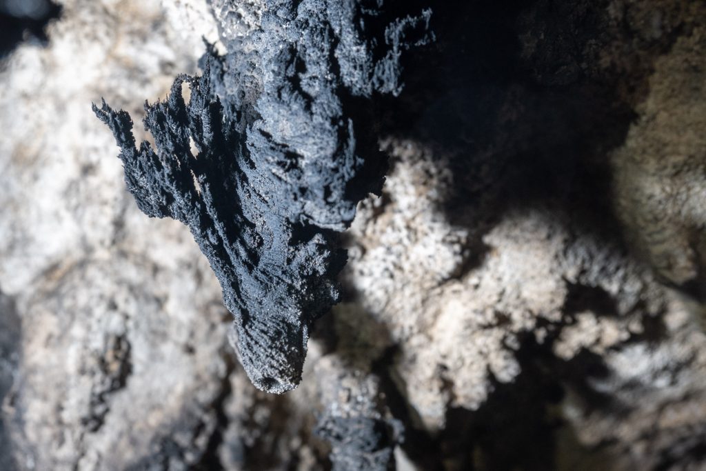
Research Area We study the interplay between the organic (microbes) and the inorganic (minerals) at the microscopic scale. Through a combination of metabolic and geochemical pathways, bacteria can initiate and influence mineral growth and/or decay. These processes are often influenced by cave climatology, creating extremely rare types of speleothems (calcium carbonate structures). Caves host a diverse range of microbial communities in close association with these speleothems, making these environments important sources of scientific discovery relating to mineralogy and geomicrobiology. We aim to investigate the mineralogy unusual speleothems collected from a remote cave and search for evidence of microbial influences in its formation. Current Project I am seeking a student interested in helping me investigate the mineralogical and microbiological aspects of cave-derived specimens using a combination of microscopy and geochemical techniques. Student will utilize a range of analysis techniques potentially including X-ray powder diffraction, Raman spectroscopy, and microscopy. Students will begin to gain a practical understanding of mineralogical research, develop a range of laboratory skills, and assist in planning and carrying out experiments.
Fig. 1: carbonate speleothem showing strong climatological influence and potentially microbial influence. Photo by Dr. Jean Krejca, Zara Environmental, LLC

Click here for more information about Dr. Barton’s lab

Do spider egg sacs protect against desiccation?

[Past Projects]

Dr. Todd Blackledge and Kate Karkosiak


Spiders’ egg sacs are woven from multiple types of spider silk and offer protection to developing embryos and spiderlings. There is extreme diversity among egg sacs across species in shape, color, texture, and habitat, and little is currently known about how these variations affect spider development.
Extreme water loss is one threat to developing spiders that can cause them to desiccate, or dry out. I am studying whether spider egg sac materials protect against desiccation of developing spiders by preventing water from escaping the inside of the egg sac. I would also like to explore which characteristics of the egg sac silk formations contribute to lower or higher transmission rates through the material.
Skills you will acquire:
  • Knowledge about spider biology and silk
  • Experience caring for invertebrate animals
  • Knowledge of materials and imaging methods
  • Field experience and sample collection
  • Research experience
  • Scientific reading/writing

Click here for more information about Dr. Blackledge’s research.

Research in Acoustic Communication and Emotions

[Past Projects]

Jeffrey Wenstrup, Ph.D., Sharad Shanbhag, Ph.D., and Mahtab Tehrani, Ph.D.


Our Work Vocalizations reflect our emotions. As we listen to another’s vocal signals, we respond by assigning them a meaning determined by the vocalization itself, our previous experience, and our own emotional state. This in turn affects the way that we respond to a person’s vocal signals—by our posture, facial gestures, movement, and speech. Our goal is to understand how the brain shapes our responses to these vocal signals.
Our approach Brain regions of interest: Our focus is on the amygdala, a brain center that integrates sensory inputs with information about our previous experiences and our internal state. It then assesses the meaning of new sensory information and “decides” on the appropriate behavioral responses. We also study the auditory centers that feed information to the amygdala about social vocalizations.
Our models We use bats and mice as models to study these processes. Bats are sound experts that use vocalizations to both communicate and to catch prey and navigate through echolocation. Mice are acoustic generalists that integrate acoustic and other sensory information during social interactions. Both models provide valuable insights into the mechanisms underlying acoustic communication and emotions. Our techniques: We record and analyze the social vocalizations of bats and mice to understand how they communicate their emotional state through social vocalizations. These analyses help us to design vocal stimuli to study the animals’ behavioral and brain responses to emotion-containing vocalizations. Our neural studies use a combination of anatomical tracing, optogenetics, imaging, electrophysiology, microdialysis, and pharmaceutical techniques. These help us to describe how amygdala neurons respond to vocalizations, which brain areas drive the amygdalar responses to vocalizations, and how modulatory neurochemicals shape these neuronal responses and behavioral reactions. Research Projects involving undergraduates • Describing vocal behavior in mice • Analyzing circuitry related to emotions and vocalizations • Analyzing neural responses to vocalizations Publications https://scholar.google.com/citations?hl=en&user=VxcRjv0AAAAJ&view_op=list_works&sortby=pubdate
Click here for more information on the Wenstrup lab.

Digital Anatomical Dissection

[Past Projects]

Dr. Rachel Olson


The Olson lab is interested in the form and function of the mammalian feeding system. We look at this through studies of mammalian diversity and evolution, as well as using animal models to better understand human disorders. A multitude of anatomical structures are involved in the feeding apparatus, including the tongue. These muscles have complex orientations that are hard to dissect and measure. Therefore, we use diceCT scanning methods, where the soft-tissues are differentially stained with an iodine-based contrast enhancing stain, to digitally dissect these structures. Digital dissection preserves the 3D orientation of structures while allowing us to visualize and quantify the structures of interest.

Primary Goals

  1. Digitally dissect the muscles involved in chewing (multiple individuals and species have been scanned).
  2. Determine the fiber orientations of the tongue.
  3. Potential to develop an analysis method to compare different species.
  4. Potential to apply anatomical data of the tongue to in vivo biomechanics of the tongue.
Skills (no experience necessary):
  • Anatomical imaging – learn about and look at CT scans and x-ray imaging. There may be opportunities to participate in collecting this data.
  • Digital dissection methods using image processing software like Slicermorph and VGstudio.
  • Interact with and learn anatomy and biomechanics in an applied context.
  • Contrast-enhanced staining methods in the wet lab.
  • Possibility of traditional gross dissections.
  • Data visualization and statistical analysis in R.
  • Participate in lab meetings and scientific discussions.
  • Participate in the Biological Undergraduate Research Symposium.

Click here for more information about Dr. Olson’s lab

The effects of invasive and naturalized worms on plant growth

[Past Projects]

Dr. Randall Mitchell and Cynthia Yoder


The Mitchell lab studies various ecological questions, from plants to animals and the human interactions with the environment. For this project, we will be studying the way worms change soil structure and how they may be affecting plants within the soils in which they exist.
In this project we will be conducting a laboratory experiment using three species of worms and produced castings to grow plants, measuring the changes in growth during this process, as well as changes in flower production and others. You will learn about the use of composting worms in agriculture and home gardening, hands-on scientific study of collecting and analyzing data, soil and worm sampling methodology, sample preparation and preservation, and applied scientific work, along with soil processes and more.
Click here for more information about the Mitchell lab.

Zebrafish as a model for bone remodeling

[Past Projects]

Dr. Richard Londraville and Kathryn Bisaha


Castranova, D., Weinstein, B., Samasa, B., n.d. Dorsal view of bones and scales (blue) and lymphatic vessels (orange) in a juvenile zebrafish | 2020 Photomicrography Competition. 
The Londraville lab seeks to explore the relationship between endospanin, leptin and bone in zebrafish through the comparison of various mutant strains. In this project students will explore how some variable (e.g., age, weight, mutation) affects bone formation and bone resorption in zebrafish. You will gain hands-on experience in the following areas: zebrafish handling and care, ImageJ analysis, microscopy, microinjection, and protein assays.
Click here for more information on Dr. Londraville’s lab.

Digital tracing neurons in a mouse model of glaucoma

[Past Projects]

Dr. Jordan Renna and Sara Chaszeyka


Our lab is studying the effects of the debilitating eye disease glaucoma on melanopsin ganglion cell neurons in the mouse retina. Melanopsin ganglion cells send light-evoked signals to the brain to regulate our circadian rhythms. Glaucoma is a medical condition in which eye pressure increases to the point at which neurons connecting the eye to the brain begin to die.   We are looking for a brave soul to digitally trace fluorescently labeled melanopsin ganglion cell neurons.  These neurons are from retinas in various stages of glaucoma. Our hope is to one day resolve glaucoma and cure the leading cause of rodent blindness. Our lab motto: NO BLIND MICE!
Click here for more information on Dr.Renna’s lab.

Synaptic Connections between light detecting cells and interneurons in the zebrafish retina

[Past Project]

Dr. Jordan Renna and Matthew Tarchick


Our lab utilizes a procedure called Electroretinogram (ERG) to study the behavioral and physiological properties of the neural retina. This protocol has medical relevance and is routinely done by optometrists. It demonstrates the whole function of the retina and the function of several important cell populations. The A wave of the ERG is related to total output of light detecting cells (rods and cones). The B-wave of the ERG is related to the total output of the interneurons (or bipolar cells). These cells form a complex synapse that is needed for the transmission of visual information.  

Our new project aims to classify how these connections are established and occur in zebrafish. Very few experiments have been done to measure the responsiveness of the zebrafish retina, so this approach is somewhat novel. The ease at which zebrafish can be genetically altered also opens more possible avenues for studying disease models and makes zebrafish a good model organism.  

We are looking for students to help run ERG experiments and conduct the analysis of those experiments.  


Click here for more information on Dr.Renna’s lab.

Biomechanics of Locomotion in an Animal Model of Preterm Infant Motor Development

[Past Projects]

Dr. Jesse Young (Department of Anatomy and Neurobiology, Northeast Ohio Medical University)


Preterm infants constitute 11% of all infants born in the United States.  Most of these infants show significant delays in motor development. However, the exact physiological mechanisms that account for these delays remain a mystery. Previous studies have hypothesized causes as disparate as early postnatal respiratory distress, poor cerebellar development, low muscle mass, and limits on muscle power production. In part, this gap in our knowledge stems from the integrative nature of locomotion itself.  Safe, efficient walking requires the coordinated output of multiple organ systems (respiratory, circulatory, nervous, musculoskeletal) to modulate the energetic and biomechanical demands of supporting and accelerating body mass. Only a longitudinal, multimodal study could provide the type of integrative physiological and mechanical data required to address the etiology of preterm infant motor delays. Such a study is not feasible in a compromised population like preterm human infants. Our lab has been developing the infant pig as an animal model of preterm human infant motor development. You would contribute to this goal by analyzing and interpreting previously collected data on walking, running, and standing in preterm and term infant pigs, contributing to our understanding of the underlying causes of preterm motor dysfunction. Skills you will develop:
  • Familiarity with modern biomechanical techniques used to analyze motor function and coordination in animals and humans
  • Use of MATLAB and R for data analysis and statistical interpretation
  • Opportunities to participate in weekly Biomechanics Journal Club meetings in the Department of Anatomy and Neurobiology at NEOMED and attend associated seminars

More about locomotor research in the Young Lab: www.younglaboratory.org.

‘I’m too freaked out to hear you!’ How early-life stress affects auditory perception and the brain

[Past Projects]

Merri Rosen PhD, Kate Hardy MS, and Matthew Sunthimer BS


It’s well-known that hearing problems in kids may cause later trouble with understanding complex sounds, such as rapid speech in a place with a lot of background noise. But our lab is the first to show that stress during development can cause similar problems. We’re funded by the National Institutes of Health to study the neural changes that can cause these stress-induced deficits in auditory perception. Many labs have studied the effects of early life stress (ELS) on the development of brain regions responsible for attention, learning, and related psychopathologies. However, nobody has examined whether auditory information is being encoded properly before it even reaches these higher-level brain regions. This is important, because children who grow up in low-socioeconomic, high stress environments are at risk for later problems with speech perception. Discovering what is going wrong mechanistically will help prevent and remediate these problems.
Our lab has shown that ELS affects auditory perception and neural activity in brain regions that encode sound. It turns out that ELS may be a particular problem when kids have ear infections that cause intermittent hearing loss – our data show that early hearing loss and stress together are much worse than either one alone! We use an animal model (the Mongolian gerbil) to study how early-life stress and hearing loss affect the perception of rapid changes in sound, and the underlying neural mechanisms. The Mongolian gerbil is a well-established auditory model, because gerbils hear well at frequencies that humans use, unlike mice or rats. Several projects are available in our lab: Seeing stress in the brain!
  • Background: Auditory perceptual problems from ELS and hearing loss may arise from changes in certain molecules that determine how neural circuits are wired during development. Measuring these elements will clarify whether deficits from ELS and hearing loss arise through similar or disparate mechanisms.
  • Objectives: To investigate changes in specific molecular markers within the auditory pathway across development. These changes are indicative of alterations in functionality and plasticity across development.
  • Methods: The effects of early stress and hearing loss on the brain will be identified using immunohistochemistry techniques which tag specific neuronal proteins with fluorescent markers. These markers can be visualized and quantified under a fluorescent microscope.
What stresses out our animals? Perception in action: Testing the limits of hearing
  • Background: Historically, mice and rats have been primarily used in stress research. To study stress in an established animal model of hearing, we are developing a new model of stress in the Mongolian gerbil.
  • Objective: To characterize the effects of the limited bedding model (LBM) on emotion, cognition, and auditory perception.
  • Methods: Implementation of the LBM model uses video recordings of breeder cages in a bedding-scarce environment. A combination of behavior tracking software and manual hand scoring of recordings will be used to characterize the new model. Various biomarkers will be measured to quantify stress markers, such as blood corticosterone and body weight. Effects on anxiety and cognition will be measured by standardized behavioral tests. Deficits in auditory perception will be measured using operant conditioning to train animals to respond when they discriminate between behaviorally-relevant sounds.
Livestreaming real-time brain activity: The Miniscope
  • Background: Our lab is working to set up an optical Miniscope, which allows us to visualize neuronal activity across a population of cells in real time, in freely-moving behaving animals.
  • Objectives: To contribute to getting this leading-edge technology set up in the lab.
  • Methods: Various skills will be learned while working closely alongside a lab member.
Benefits to student:
  • Receive training in experimental techniques used widely across research fields in biology and neuroscience
  • Learn how to apply critical thinking to big problems in neuroscience
  • Opportunity to join our lab as a PhD student or technician

Learn more about the Rosen lab.