[Past Projects]
Dr. Jordan Renna and Katelyn Sondereker
In general, our lab studies the eye: We study the retina and how it develops. More specifically, we are interested in the development of melanopsin ganglion cells. Melanopsin ganglion cells are like rods and cones, but they send the brain non-visual information about circadian rhythms and the pupillary light reflex. Melanopsin ganglion cells are also a target for studies involving health and disease, since they are resistant to many visual disorders.
My current project involves studying the morphology of melanopsin ganglion cells in a mouse model of glaucoma. Staining retinal tissue to visualize melanopsin will allow us to determine how the anatomy of melanopsin ganglion cells changes as the disease progresses. This will provide invaluable information about the effect of glaucoma on melanopsin ganglion cells. I am looking for students to become independent in their ability to perform eye dissections (Fig. 1, 2), stain tissue with immunohistochemistry (Fig. 3), and trace the morphology of melanopsin ganglion cells with programs like ImageJ (Fig. 4). These techniques will be useful to anyone interested in a career in the medical field or in biomedical and clinical research.
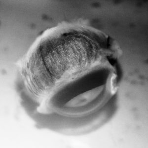
Figure 1: A lateral view of an enucleated mouse eye before dissection.
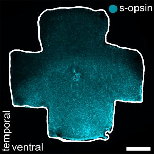
Figure 2: An isolated mouse retina immunostained for s-opsin (blue).
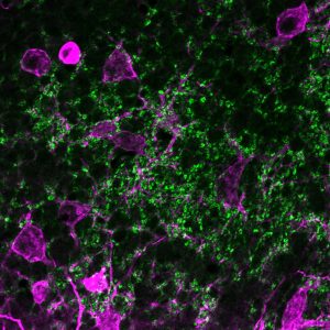
Figure 3: Retinal tissue immunostained for melanopsin (magenta) and VGLUT2 (green).
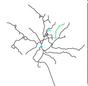
Figure 4: A whole-cell reconstruction tracing of a melanopsin ganglion cell.