[Past Projects]
Dr. Janna Andronowski and Reed Davis
The misuse and addiction to opioids (and synthetic opioids) is a serious public health crisis nationwide that has become an epidemic. Current evidence suggests that opioids upset the balance of bone remodeling towards more destruction and less formation of bone. Experimental studies have been limited by the fact that small laboratory animals traditionally used in bone research (mice and rats) do not exhibit spontaneous cortical bone remodeling, making them a poor choice of animal model for this subject.
A new project in the Andronowski Lab seeks to develop a long-term model for studying the effects of prolonged opioid use on cortical bone remodeling in an animal which remodels its cortical bone in a manner comparable to humans, the rabbit. An innovative 3D X-ray imaging technique (micro-CT), combined with dynamic histomorphometry, will allow us to describe how morphine and fentanyl affect microscopic structures of cortical bone used in histological age estimation methods in forensic anthropology. Given the limited data available related to the impact of opioid abuse on bone remodeling, our goal is to further understandings of the underlying biological processes and improve the applicability of histological age-estimation methods and scientific standards within the field of forensic anthropology.
Prospective students can expect to learn about how we apply the principles of bone remodeling to estimate age-at-death in forensic anthropology, 3D X-ray imaging using desktop micro-CT (Figure 3), big data processing, the preparation of bone tissue slides, animal handling, and dissection techniques. Dr. Andronowski is interested in undergraduate students from any major who are willing to learn the above skills. Preference will be given, however, to those who have experience with live animal research, animal dissections, histological techniques, and computer programming/modeling.
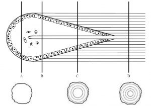
Figure 1: Coupled osteoclast and osteoblast activity depicted as a Basic Multicellular Unit during bone remodeling. A) Osteoclastic activity near the leading edge of the cutting cone, B) initiation of osteoblastic activity, C) active osteoblast activity, and D) a fully formed intact osteon depicting a Haversian Canal in the center.
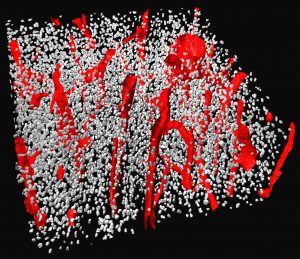
Figure 2: Micro-CT 3D render of cortical bone showing vascular porosity (red) and osteocyte lacunae (grey). Data collected at the Canadian Light Source synchrotron.
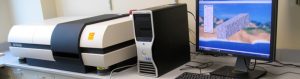
Figure 3: SkyScan 1172 Desktop Micro-CT system at the University of Akron.