Evaluating Postmortem Changes to Human Bone Microstructure Using Virtual Histology
[Past Projects]
Dr. Janna Andronowski and Randi Depp
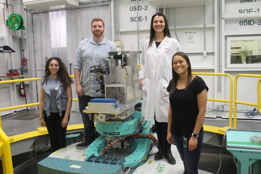
Figure 1: Andronowski Lab ‘Beam Team’ members during experiments at the Canadian Light Source national synchrotron facility in Saskatchewan, Canada. From left to right: Former UA undergraduate Linda Muakkassa, UA doctoral student Reed Davis, Dr. Andronowski, and former UA undergraduate Hannah Stephen.
What do forensic science, bones, X-ray beams, and 3D imaging have in common? The Andronowski Lab!
Research background:
The estimation of the postmortem interval (PMI), or time since death, is a crucial and fundamental consideration in forensic death scene investigations. With the progression of time and soft tissue decomposition, a postmortem estimation by a medical examiner becomes more difficult and less accurate, as bone and teeth are often the only remaining tissues of an organism. Diagenetic agents (e.g., microbial infiltration, ground water, soil, pH balance, fungi) are known to structurally alter bone tissue micromorphology over time. This may further disrupt high-quality DNA samples or histological age-at-death estimates in forensic anthropological contexts. To date, there are no quantitative tools available to the forensic anthropologist to evaluate bone diagenesis.
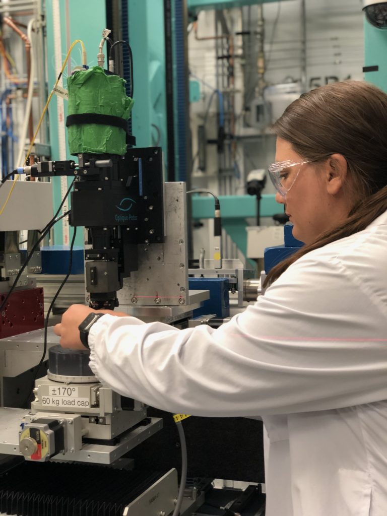
Figure 2: Current Tiered Mentoring student, Gina Tubo, during experiments at the Canadian Light Source.Project details:
Goal: To identify and quantify bone tissue microarchitectural changes across increasing postmortem intervals and evaluate how various diagenetic agents impact microstructures of cortical bone used in histological age estimation methods in forensic anthropology.
Methods: An innovative 3D X-ray imaging technique (Synchrotron Radiation-based micro-CT) will be utilized. High-resolution imaging experiments will be executed at the Canadian Light Source national synchrotron facility.
Sample groups: Four predetermined postmortem intervals (0-3 years, 4-10 years, 11-20 years and >20 years) were identified and three skeletons selected to represent each interval (n=12).
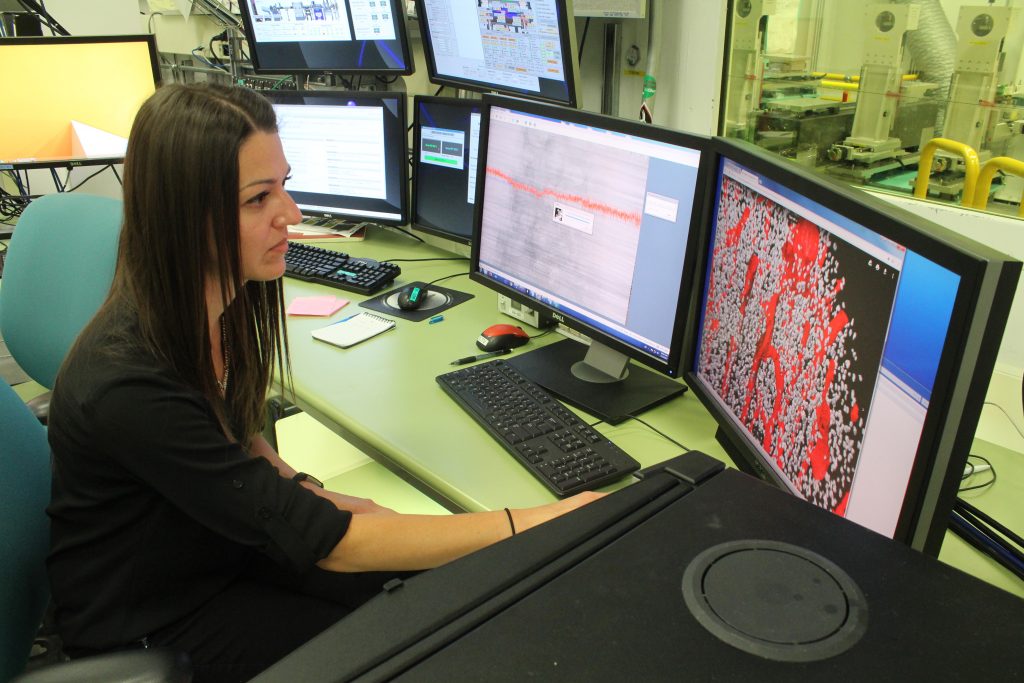
Figure 3: Dr. Andronowski visualizing cortical bone data at the Canadian Light Source.Benefits for the Student:
Learn about how we apply the principles of bone remodeling to estimate age-at-death in forensic anthropology
Receive training in 3D X-ray imaging using desktop micro-CT, big data processing, the preparation of bone tissue slides, and dissection techniques.
As a Tiered Mentoring student, you may have the opportunity to join the Andronowski Lab ‘Beam Team’ and assist with imaging experiments for this project.
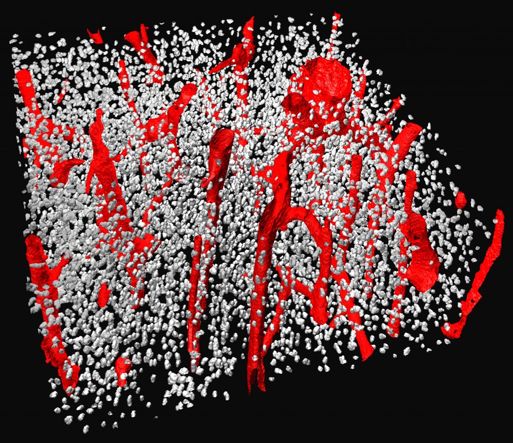
Figure 4: 3D render of cortical bone showing vascular porosity (red) and bone cellular spaces (grey). Data collected at the Canadian Light Source.Qualifications:
Dr. Andronowski is interested in undergraduate students from any major who are willing to learn the above skills. Preference will be given, however, to those who have experience with dissection (animal and/or human), histological techniques, and computer programming/modeling.
Click here to learn more about Dr. Andronowski’s lab