Using Robots to Understand Fish Maneuverability

[Past Projects]

Dr. Henry Astley and Stephen Howe


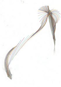
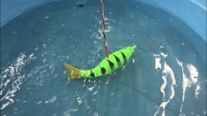
Fish are adept swimmers that control their locomotion using relatively simple methods. We have been gathering swimming data from live fish and creating models to describe their behaviors. Working with live fish comes with certain limitations due to unpredictable fish behavior and the inaccessibility of rare or delicate species. We are developing a robotic model to test hypotheses about turning behavior in fish that we would not be able to test with live fish. This robot will help us understand how fish control turns and what are the limitations of their maneuverability. The robot will allow us to study the relationship between body shape and swimming performance to add some perspective to a long-standing debate in the fish swimming literature. Expect to learn about robotics, 3D modeling, videography and image tracking, and fish biomechanics. We are interested in undergraduate students from any major that are willing to learn any of the above skills. No prior experience is necessary.


Click here to learn more about the Astley Lab


Sticky When Wet: Understanding Tree Frog Adhesion on Different Surfaces

[Past Projects]

Dr. Henry Astley, Dr. Peter Niewiarowski, Derek Jurestovsky, and Austin Garner


Research Description

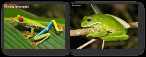
Tree frogs secrete a mucous on the bottom of their micropatterned toe pads that permit them to adhere to surfaces like two wet glass microscope slides stuck together (capillary adhesion). Theoretically, tree frog adhesion should not be dependent on whether the surface of interest repels or attracts water because the secreted mucous is capable of sustaining adhesive contact on a variety of surfaces. In this study, we will measure live tree frog adhesion under dry, misted with water, and wet conditions using a variety of artificial surfaces that range in their water repellency.

Benefits of this Research Experience

Undergraduate students in the Astley and Niewiarowski labs will gain an array of critical research skills including: amphibian care and handling, live animal performance measurements, experimental design, statistics, microscopy, surface characterization methods, scientific writing, and more. Additionally, many of the undergraduate students in our labs have been co-authors on several papers published in peer-reviewed journals (see below)!


*Denotes undergraduate student

Niewiarowski, P. H., Lopez, S., Ge, L., Hagan, E.* and Dhinojwala, A. (2008). Sticky gecko feet: the role of temperature and humidity. PLoS. ONE 3, e2192.

Niewiarowski, P. H., Stark, A., McClung, B.*, Chambers, B.* and Sullivan, T.* (2012). Faster but Not Stickier: Invasive House Geckos Can Out-Sprint Resident Mournful Geckos in Moorea, French Polynesia. J. Herpetol. 46, 194-197.

Stark, A. Y., Sullivan, T. W.* and Niewiarowski, P. H. (2012). The effect of surface water and wetting on gecko adhesion. J. Exp. Biol. 215, 3080-6.

Stark, A. Y., Badge, I., Wucinich, N. A.*, Sullivan, T. W.*, Niewiarowski, P. H. and Dhinojwala, A. (2013). Surface wettability plays a significant role in gecko adhesion underwater. Proc Natl Acad Sci U S A 110, 6340-5.

Stark, A. Y., Wucinich, N. A.*, Paoloni, E. L.*, Niewiarowski, P. H. and Dhinojwala, A. (2014). Self-drying: a gecko’s innate ability to remove water from wet toe pads. PLoS ONE 9, e101885.

Stark, A. Y., McClung, B.*, Niewiarowski, P. H. and Dhinojwala, A. (2014). Reduction of water surface tension significantly impacts gecko adhesion underwater. Integr. Comp. Biol 54, 1026-33.

Badge, I., Stark, A. Y., Paoloni, E. L.*, Niewiarowski, P. H. and Dhinojwala, A. (2014). The role of surface chemistry in adhesion and wetting of gecko toe pads. Scientific Reports 4, 6643.

Stark, A. Y., Ohlemacher, J.*, Knight, A.* and Niewiarowski, P. H. (2015). Run don’t walk: locomotor performance of geckos on wet substrates. J. Exp. Biol. 218, 2435-41.

Stark, A. Y., Palecek, A. M.*, Argenbright, C. W., Bernard, C.*, Brennan, A. B., Niewiarowski, P. H. and Dhinojwala, A. (2015). Gecko adhesion on wet and dry patterned substrates. PLoS. ONE 10, e0145756.

Stark, A. Y., Dryden, D. M., Olderman, J.*, Peterson, K. A., Niewiarowski, P. H., French, R. H. and Dhinojwala, A. (2015). Adhesive interactions of geckos with wet and dry fluoropolymer substrates. J. R. Soc. Interface 12, 20150464.

Stark, A. Y., Subarajan, S.*, Jain, D., Niewiarowski, P. H. and Dhinojwala, A. (2016). Superhydrophobicity of the gecko toe pad: biological optimization versus laboratory maximization. Phil. Trans. R. Soc. A 374, 20160184.

Klittich, M. R., Wilson, M. C., Bernard, C.*, Rodrigo, R. M.*, Keith, A. J.*, Niewiarowski, P. H. and Dhinojwala, A. (2017). Influence of substrate modulus on gecko adhesion. Scientific Reports 7, 43647.

Garner, A. M.*, Stark, A. Y., Thomas, S. A. and Niewiarowski, P. H. (2017). Geckos go the Distance: Water’s Effect on the Speed of Adhesive Locomotion in Geckos. J. Herpetol. 51, 240-244.


Click here to learn more about the Astley Lab

Click here to learn more about the Niewiarowski lab
Investigating the morphology of melanopsin ganglion cells in health and disease

[Past Projects]

Dr. Jordan Renna and Katelyn Sondereker


In general, our lab studies the eye: We study the retina and how it develops. More specifically, we are interested in the development of melanopsin ganglion cells. Melanopsin ganglion cells are like rods and cones, but they send the brain non-visual information about circadian rhythms and the pupillary light reflex. Melanopsin ganglion cells are also a target for studies involving health and disease, since they are resistant to many visual disorders.

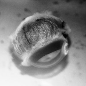
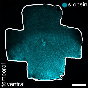
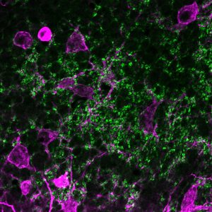
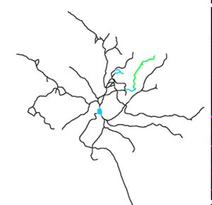
My current project involves studying the morphology of melanopsin ganglion cells in a mouse model of glaucoma. Staining retinal tissue to visualize melanopsin will allow us to determine how the anatomy of melanopsin ganglion cells changes as the disease progresses. This will provide invaluable information about the effect of glaucoma on melanopsin ganglion cells. I am looking for students to become independent in their ability to perform eye dissections (Fig. 1, 2), stain tissue with immunohistochemistry (Fig. 3), and trace the morphology of melanopsin ganglion cells with programs like ImageJ (Fig. 4). These techniques will be useful to anyone interested in a career in the medical field or in biomedical and clinical research.


Figure 1: A lateral view of an enucleated mouse eye before dissection.


Figure 2: An isolated mouse retina immunostained for s-opsin (blue).


Figure 3: Retinal tissue immunostained for melanopsin (magenta) and VGLUT2 (green).


Figure 4: A whole-cell reconstruction tracing of a melanopsin ganglion cell.


Click here for more information on Dr.Renna’s lab.

Macro-invertebrates 3D modeling

[Past Projects]

Dr. Francisco Moore and Banafsheh Khakipoor


Do you aspire to come up with creative ways to teach youth? Are you excited about 3D printers? If yes, then consider applying for this project.

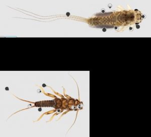
This project is a collaboration with Cleveland Metro Parks to develop a K-12 curriculum for water quality monitoring by learning about macro-invertebrates. We create three-dimensional models of the critters living in a watershed to use as a teaching tool. This is especially beneficial when students cannot go to a water body to study these invertebrates in their habitats due to weather, accessibility, etc. An indoor classroom can mimic a river outline and use the 3D models as replicas.

In this project we will use and learn:

  • Collect Macro-invertebrates from Watershed Stewardship Center
  • Use a Micro CT scanner to scan these marco-invertebrates.
  • Use ImageJ (image analysis software) to create 3D models using series of images taken by MicroCT scanner.
  • Print the models using a 3D printer.

Click here for more information on Dr. Moore’s lab.

Tracking human health & diet with stable isotopes

[Past Projects]

Dr. Anne Wiley and Nate Michael


Could you tell what a stranger ate during the past month, using only a tiny sample of their hair? Could you predict their risk of diabetes, hypertension, or other chronic disease? These are the questions at the heart of our research… and potentially at the heart of your new project.

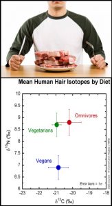
Unbeknownst to most people, our dietary history is recorded by subtle shifts in the chemical composition of our hair. Stable isotopes are alternative forms of an element that differ in their number of neutrons (e.g. 14N has 7 neutrons while 15N has 8); the ratio of these isotopes in human tissues are inherited from our food. For example, people who consume meat and other animal-based foods have more 15N in their hair than vegans. And the more corn syrup people consume, the more 13C will become concentrated in their blood. Because stable isotopes reflect aspects of diet such as sugar consumption that are linked with obesity and chronic disease, many researchers have predicted that they will have a powerful impact in future human health studies.

The goal of this project is to collect hair samples from University of Akron students and determine exactly what aspects of diet can (and can’t) be quantified using stable isotope techniques.  We’re searching for a dedicated undergraduate who can help to collect hair samples from volunteers, help administer diet and health surveys, learn to analyze samples for their isotopic content in lab, and push our understanding about the isotope-diet link to the next level. The recruited student will join an interdisciplinary team (ecologists and stable isotope specialists, an anthropologist, and an expert in human movement) who are aiming to apply stable isotopes to the study of food availability and health in low-income Cleveland neighborhoods. The student may have the opportunity to join in the Cleveland-based research, depending on their timeline.


Click here to learn more about Dr. Wiley’s lab.

Classifying Ion Channels in the Retina

[Past Projects]

Dr. Jordan Renna and Matthew Tarchick


Our lab studies the development of circuits in the retina. During development, an interconnected layer of the retina called the starburst amacrine cell layer fires bursting patterns. These patterns occur for 10-12 days. A specific interest to me is the how potassium and calcium conductance plays a modulating role in these bursting patterns, and how those bursts can pass on information. To better understand conductance, we are exploring the expression of ion channel proteins and genes, as well as visualizing their location, and using agents which inhibit those channel activities.

To study this, we use many typical molecular biology methods like immunohistochemistry, western blotting and quantitative PCR. With western blotting we can get an idea of the change in protein expression and monitor the presence of a protein in the retina. With Immunohistochemistry we can get a better look at the localization of a certain protein. I am looking for students that can help me by learning these techniques. These techniques are very valuable for careers in medical research and various other biomedical studies.


Fig 1. Retinal Lysate Western Blot of Beta Actin


Fig 2. Immunohistochemistry:

Blue is DAPI, a nuclear stain

Red is choline acetyltransferase

Green is small conductance potassium channel 1


Click here for more information on Dr.Renna’s lab.

Bird nest construction for biomimetic insight

[Past Projects]

Dr. Hunter King and Meron Dibia


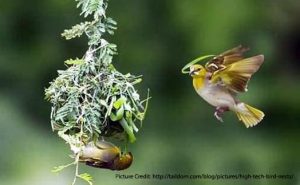
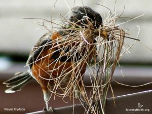
This project will examine the material selection for and construction protocol of a bird’s nest. We are exploring the material science responsible for the highly tuned structural properties of nests, despite their use of seemingly random and flimsy raw materials.


In order to get more data from the construction of the nest, we will be collaborating with Akron Zoo to monitor the nesting behavior of a couple of species in captivation.

You will be helping with monitoring and maintaining the cameras at the zoo, and examining video footage to extract usable data. We will accept an undergraduate from any major who is comfortable using photo/video equipment and interested in custom field instrumentation.

(Spring Semester, 2019)


Click here for other information about Dr. King.

Click her for more information about projects in Dr. King’s lab.

Bio-inspired, passive vapor harvesting with thermoresponsive polymer fibers

[Past Projects]

Dr. Hunter King and Aida Shahrokhian


Today, about one billion people worldwide lack access to safe water. This global demand for fresh water is projected to increase as global population rises. Water in the form of vapor and droplets within the atmosphere is estimated to be about 13 thousand trillion liters, which makes it a good candidate for drought-prone and water-scarce areas. Many plant and animals such as beetles, frogs, spiders, Opuntia microdasys, Stipagrostis sabulicola, and Trianthema hereroensis have evolved to benefit from this atmospheric moisture, where there is no other source of water available. There have been many attempts to mimic these systems to harvest water from the air in the past few decades.


Our motivation for this project is to create a passive system where water vapor is absorbed at a certain condition and then released in another without applying any energy. For this application, various biomimetic and synthetic materials such as spider glue salts and stimuli-responsive polymers, specifically thermo-responsive polymers with LCST are considered. Thermo-responsive polymers with LCST, have tendency to remain soluble below a particular temperature and above this temperature they undergo a change in their solubility behavior and phase separate. To investigate these materials for water harvesting application, we electrospin them and test their water collection and release behavior. In this project, your task will be to find the optimum parameters for electrospinning cellulose acetate, and to find the best curing temperature for crosslinking of electrospun PNIPAM nanofibers. Techniques you will be learning are electrospinning, Scanning Electron Microscopy(SEM), optical microscopy, Differential Scanning Calorimetry (DSC) and Fourier-transform infrared spectroscopy (FTIR).


Click here for other information about Dr. King.

Click her for more information about projects in Dr. King’s lab.

DIY Spectrometry mobile phone application development project

[Past Projects]

Dr. Hunter King and Bana Khakipoor


Mobile phones have become popular in everything we do, from photography to organizing our schedules, to science!

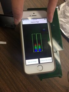
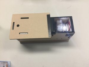
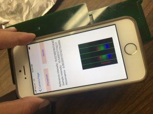
In this project we use mobile phones to do science by creating a citizen science application to measure nutrient loading in lake Erie’s tributaries. Normally one need to use a colorimeter or spectrometer to measure phosphate or nitrate concentration in water. Colorimeter costs ranges from >$1k to $3,$4k, making it inaccessible for average citizen scientists. Hence, in this project we developed smartphones + Fab Lab produced spectrometers to hack the spectrometer industry!


You’ll be using Xamarin to develop our application for Android, Windows, and iOS platforms. We already have done some works in Xamarin.iOS which could be your starting point.  You can either be a developer or a designer. Our developer needs to be computer science undergraduate. Our designer needs to be innovative and think of how this technology can become more inviting and intriguing for citizen scientists.


Click here for other information about Dr. King’s lab.

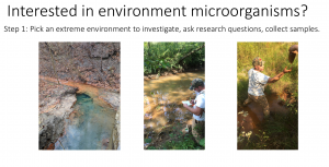
Can we use microorganisms in nature to solve environmentally related problems like acid mine drainage (AMD)?

[Past Projects]

Dr. John Senko and Shagun Sharma


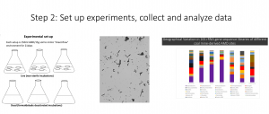
One of the project in our lab is investigating the role of microbial communities associated with acid mine drainage (AMD) in bioremediation. Coal-mine derived acid mine drainage (AMD) is formed upon intrusion of oxygenated water into the abandoned mine works and waste rock, inducing microbially mediated oxidation of FeS phases, and yielding acidic fluids with high concentrations of Fe(II). Which eventually precipitates as Fe(III) (hydr)oxides forming “yellow boys’, which smother stream substrate and destroy benthic communities.

Benefits for the students

  • Understanding of advanced molecular techniques
  • Genomics experience (Bioinformatics tools)
  • Learn to use integrated approaches to solve research questions
  • Opportunity to attend scientific conferences and building networks


Click here to learn more about Dr. Senkos’ lab.