1. Noninvasive monitoring of tumor hypoxia using FD-NIRS
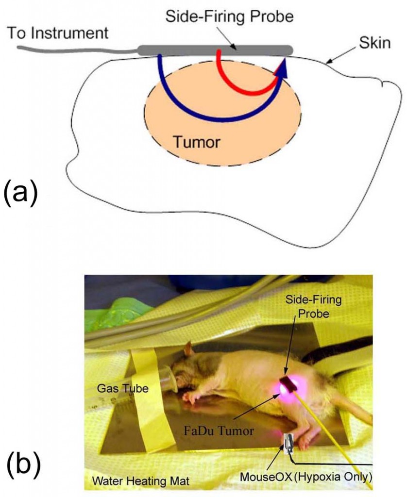
Numerous studies have found that hypoxia (lack of oxygen) leads to enhanced tumor metastasis and resistance to radiation and drugs. Strategies to reduce hypoxia by increasing oxygen delivery and decreasing oxygen consumption within the tumor are both being explored to overcome hypoxia-induced resistance. Thus there is a growing demand for technologies that noninvasively measure tumor hypoxia to enable advances in therapeutic drug development and trials. We have recently developed a side-firing fiber optic probe based on frequency-domain near-infrared spectroscopy (FD-NIRS) to quantify tissue hemoglobin content (THb) and oxygenation level (SO2) noninvasively. FD-NIRS uses RF modulated red and near-infrared lasers as the light source and a phase sensitive detection system. It measures the amplitude attenuation and phase-shift of the diffuse reflectance signal from the tissue relative to the source. A diffusion model of light transport in tissue is used to extract the tissue absorption and scattering properties from the measured reflectance spectra. The sensor can be easily and reliably attached to a tumor surface for continuous and uninterrupted measurement, and thus is an ideal tool for tumor hypoxia monitoring in vivo. The sensor has successfully captured forced and natural cycling hypoxia in FaDu rat tumor models.

 Fig. 1. Measurement of tumor hypoxia: side-firing probe attached to a FaDu rat tumor (a) and (b); tumor cycling hypoxia measured measured when the rat breathed forced cycling hypoxic gas (O2: 30%->12%->30%->12%->30%) (c) or room air (d).
2. Smart fiber optics sensor for epithelial cancer detection in LMICs
Epithelial cancers, such as oral and cervical cancers, if detected at early stages, have a better chance of being successfully treated with surgery, radiation, chemotherapy, or a combination of the three. However, in low- and middle-income countries (LMICs) epithelial cancer related death is disproportionately high due to the lack of appropriate medical infrastructure and resources to support the organized screening and diagnostic programs that are available in the developed world. Diffuse reflectance spectroscopy in the visible wavelength range (VIS-DRS) with a fiber optic probe can noninvasively quantify the optical properties of epithelial tissues and has shown the potential as a cost-effective, fast and sensitive tool for diagnosis of early precancerous changes in the cervix and oral cavity. However, current DRS systems are susceptible to several sources of systematic and random errors, such as uncontrolled probe-to-tissue pressure and lack of a real-time calibration that can significantly impair the measurement accuracy, reliability and validity of this technology as well as its clinical utility. In addition, such systems use bulky, high power and expensive optical components which impede their widespread use in LMICs.
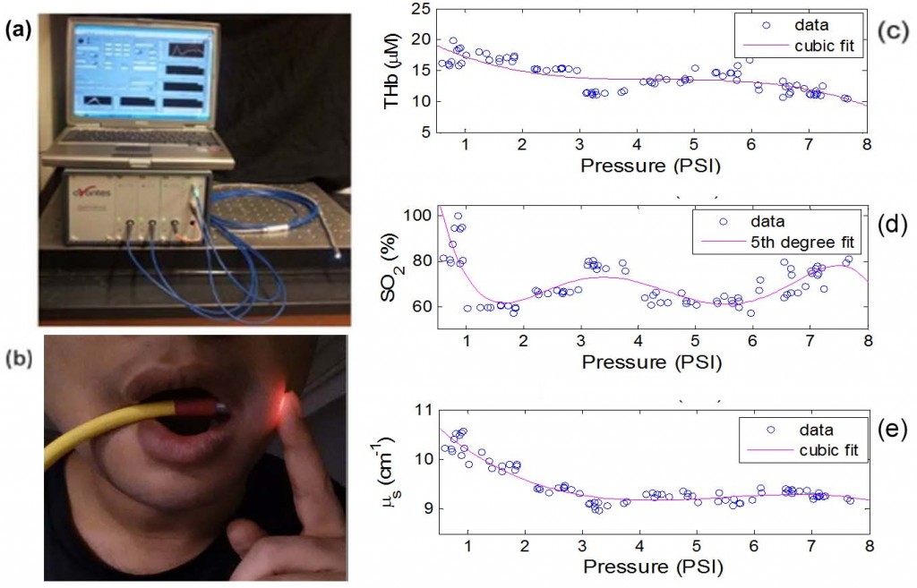
We have developed a portable, easy-to-use and low cost, yet accurate and reliable VIS-DRS device that can aid in the screening and diagnosis of epithelial cancers at an early stage in LMICs. The device integrates a VIS-DRS channel, an interferometric pressure sensor and a self-calibration channel into a singe fiber optic probe to obtain consistent DRS measurements and intact tissue optical properties. It also uses state-of-the-art photonics components to reduce power consumption and automated software to minimize the need of operator training. The device showed improved accuracy for measuring tissue scattering in both phantoms and in vivo cervical tissues. A clinical study on healthy volunteers indicated that probe pressure can significantly change tissue physiological and morphological properties and a pressure below 1.0 psi is desired for oral mucosal tissues to minimize the probe effects.

Fig. 2. (a) smart fiber optic sensor system; (b) measurement of oral tissue in a healthy volunteer; and (c-e) measured responses of healthy oral mucosa to probe pressure.
3. Monitoring of tissue damage during thermal ablation of tumors
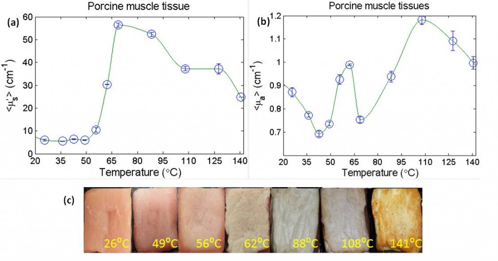
Real-time monitoring of tissue status during thermal ablation of tumors is critical to ensure complete destruction of tumor mass while avoiding tissue charring and excessive damage to normal tissue. Currently, magnetic resonance imaging (MRI) and thermometry (MRT) are commonly used for monitoring the thermal ablation process in soft tissues. Although, MRT can provide tissue damage information at the treatment site using Arrhenius damage integral, it is expensive, bulky and often subject to motion artifacts. On the other hand, light propagation within tissue is sensitive to changes in tissue microstructure and physiology which could be used to directly quantify the extent of tissue damage. Furthermore, optical monitoring can be a portable and cost-effective alternative for monitoring a thermal ablation process. In this project, we have developed a needle-based fiber-optic probe based on diffuse reflectance spectroscopy in the visible range (VIS-DRS) to quantify the optical properties during heating.
 4. Diffuse reflectance imaging of breast tumor margins
Quantitative diffuse reflectance imaging (QDRI) is an emerging modality that collects and analyzes reflectance spectra produced as ultraviolet-visible (UV-VIS) light propagated through a turbid medium to determine the absorption and scattering properties of the medium. From the absorption spectra, tissue composition maps, such the concentrations of oxy-hemoglobin (HbO2) and deoxy-hemoglobin (Hb), can be extracted, while the scattering map reflects the tissue morphological information, such as nuclear size and density. Both tissue compositions and morphological information have been identified as useful biomarkers for cancer diagnostics. We have developed a 49-channel QDRI system that can image a 4.2 cmx4.2 cm tumor margin in less than 5 seconds.
Fig. 3. QDRI for intraoperative imaging ofbreast tumor margins: (a) 49-ch QDRI probe placed on a sliced mastectomy specimen; (b) tissue total hemoglobin concentration (THb) and (c) mean reduced scattering coefficient measured from an ex vivo sample.