Background:
Banded iron formations (BIF) are the world’s largest and most widespread source of iron. The Carajas BIF of Brazil is associated with the presence of a high-grade iron ore and extensive cave development. The formation of these caves appears to be through microbial iron reduction and silica mobilization. The role and importance of silica in the formation of these caves is still unknown, so that is what we are trying to uncover.
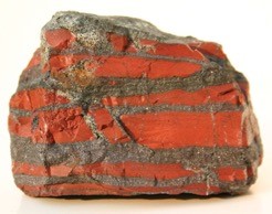
Figure 1: Banded iron formation (BIF) sample. https://kids.britannica.com/students/assembly/view/107856.Current Research Projects:
We are studying the process of silica mobilization through microbially driven iron redox reactions and the effects dissolved silica have on iron redox reactions. We use bacterial cultures grown in both anaerobic and aerobic conditions with iron and silica. We then analyze these cultures with geochemical and biological analyses.
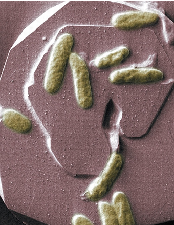
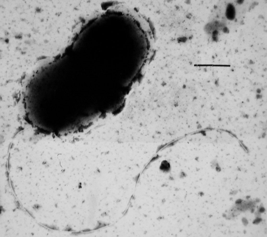
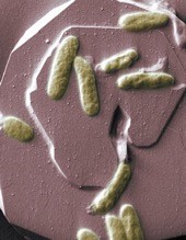
Figure 2: Shewanella oneidensis MR-1 growing on hematite, using iron as an electron acceptor. https://www.livescience.com/28163-bio-batteries-one-step-closer.htmlSkills you will develop:
Culturing bacteria · Growing bacteria in anaerobic conditions and using an anaerobic chamber · Creating bacterial growth media · Methods of geochemical analyses · Using a centrifuge, autoclave, pH meter, pipettes, spectrometer, ion chromatographer, microscope · You will begin to gain an understanding of the complex relationships between microbial life and geological processes.
Click here to learn more about Dr. Senkos’ lab.
Biomimicry is an interdisciplinary design process in which biologists become an active member of a design team, exploring biological strategies to inform innovative and potentially sustainable design solutions. In a world in which sustainable design has taken a front seat across many industries the potential of biomimicry is huge. Yet access to specific design tools and biologists within these contexts is limited.
To promote the adoption and implementation of biomimicry for sustainability within these environments this research is focused on three core areas:
1) Integrating training opportunities to promote a sustainable mindset
2) Promoting the creation of sustainable products and brand through innovation workshops within academic, industrial, and natural settings.
3) Creating cross-institutions collaborations between industries and biological institutions, Zoos and Natural History Museums, to drive scaled impact.
You will gain skills and exposure in one or more of the following:
Biomimicry design thinking methodology and tools
Curriculum design & development for environmental sustainability
Education and workshop facilitation experience with academic and industrial audiences, potentially settings.
Exposure to a variety of qualitative and quantitative social science research skills from online surveys to in-depth interview techniques and data analysis.
Interdisciplinary communication and networking skills across disciplines such as biology, engineering, design, and psychology.
If you have an interest in the field of biomimicry and its potential to promote environmental sustainability, then this is a great opportunity for you. Any undergraduate student from any major who is willing to learn and explore is invited to apply. Please note that given the current situation regarding coronavirus much of this work can be completed virtually and all measures will be taken to ensure an effective and enjoyable learning experience for all.
For more information here is a newsletter that shine some light on the type of work that has been done recently at the Cleveland Museum of Natural History. If this has caught your attention please feel free to reach out to Sarah McInernery at ssm70@zips.uakron.edu.
Click here to learn more about the Niewiarowski lab
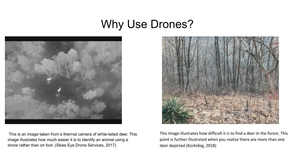
This study will use a thermal camera mounted to an unmanned aerial vehicle (UAV) to study white-tailed deer populations and migration patterns in conjunction with Bath Nature Preserve and the Summit Metroparks. While drones are a big part of the study, other methods for mapping deer will be utilized as to have something to compare the drone sampling method to.
Why Use a Drone?
The main advantages of this method of sampling is that it should be less prone to human error. Instead of a compass, protractor and rangefinder that are required for distance sampling (them method most commonly used), the drone can collect all the necessary data instantly and more reliably. Deer have developed a camouflage that makes it difficult to locate them in some environments. The thermal camera above the tree line with its line of sight not inhibited by trees take away any doubts that a researcher may have in locating and identifying the white-tailed deer. Also, the locations will be georeferenced and digitized and in GIS software any time after the fact.
For more information on this project email Stuart Davis at spd34@zips.uakron.edu.
Click here for other information about Dr. Mitchell’s lab.
For millions of years, Lechuguilla Cave has been geologically isolated, and the microbes living within it have had little to no exposure to the surface. As a result, the microbes in the cave have had to develop unusual metabolic and competitive strategies. The goal of this research is to cultivate the microorganisms that live within Lechuguilla Cave and study their adaptations to the cave environment. We will explore these adaptations by investigating methods to increase cultivation efficiency, screening isolated strains for antibiotic production, and studying the metabolism of strains that we believe are involved with biomineralizing barite (barium sulfate). Overall, this project aims to increase our understanding of the microbial ecology of Lechuguilla Cave and its potential for novel metabolisms and antibiotics.
Click here for more information about Dr. Barton’s lab
Our lab studies the development of circuits in the retina. During development, an interconnected layer of the retina called the starburst amacrine cell layer fires bursting patterns. These patterns occur for 10-12 days. A specific interest to me is the how potassium and calcium conductance plays a modulating role in these bursting patterns, and how those bursts can pass on information. To better understand conductance, we are exploring the expression of ion channel proteins and genes, as well as visualizing their location, and using agents which inhibit those channel activities.
To study this, we use many typical molecular biology methods like immunohistochemistry, western blotting and quantitative PCR. With western blotting we can get an idea of the change in protein expression and monitor the presence of a protein in the retina. With Immunohistochemistry we can get a better look at the localization of a certain protein. I am looking for students that can help me by learning these techniques. These techniques are very valuable for careers in medical research and various other biomedical studies.
Fig 1. Retinal Lysate Western Blot of Beta ActinFig 2. Immunohistochemistry:
Blut DAPI
Red Choline Acetyl Transferase
Green Small Conductance Potassium ChannelClick here for more information on Dr.Renna’s lab.
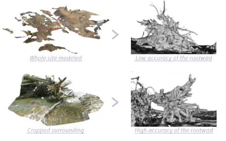
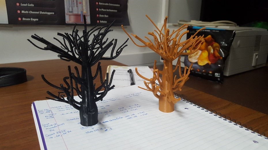
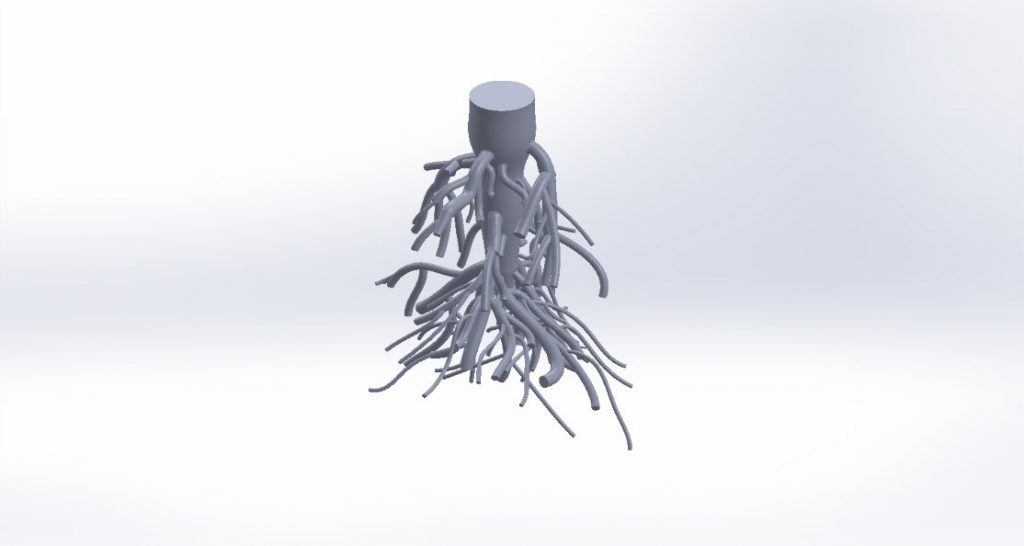
In the Biodesign Lab, we are studying the ability to design, prototype and evaluate biomimetic innovations to restore natural habitat complexity at Lake Erie. We are testing complex root shapes mimicked after native coastal forests in the region. Other root forms may be considered and explored, such as those mimicked after mangroves or coastal wetland plants.
We create 3D root models using photogrammetry and a variety of 3D software programs. We 3D print these roots using different materials for materiality and constructability exploration. Possible lab visits to Cleveland State University and Kent State University to conduct lab-scale wave attenuation and sediment depositional prediction studies may be arranged. Field work will be done in the spring to obtain adequate rootwads for imaging. The goal of the project is to contribute to the understanding of the functionality of root systems for soil stabilization through imaging, 3D information and analysis of root traits. This knowledge is then translated to innovative structural designs for coastal protection that are prototyped and evaluated using the above experimental tests.
3D models developed via Autodesk ReCap Photo from images taken in the field in March-April 2019
You’ll gain skills and exposure in one or more of the following:
Structure from motion (SfM) photogrammetry
3D modeling and printing
Materials investigation
Communication skills with a diverse array of disciplines: engineers, architects, biologists and designers
We hope students will have prior experience with 3D modeling and design, but this is not a requirement to apply. We are interested in undergraduate students from any major that are willing to learn and explore. Work at the interface of engineering, design and biology to improve the coastal ecology of Lake Erie with a Biomimicry Fellow.
3D printed PLA root structures from UA Makerspace
White oak Solidworks root model – from Liang T, Knappett JA, et al. 2017 paper in LandslidesAdditional Background:
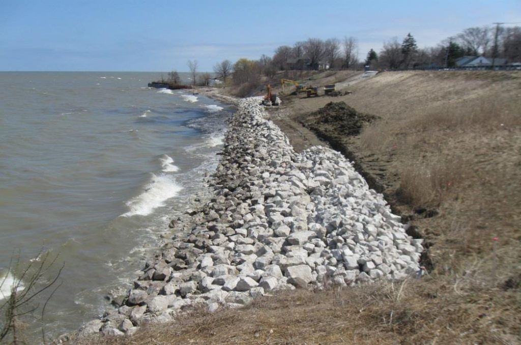
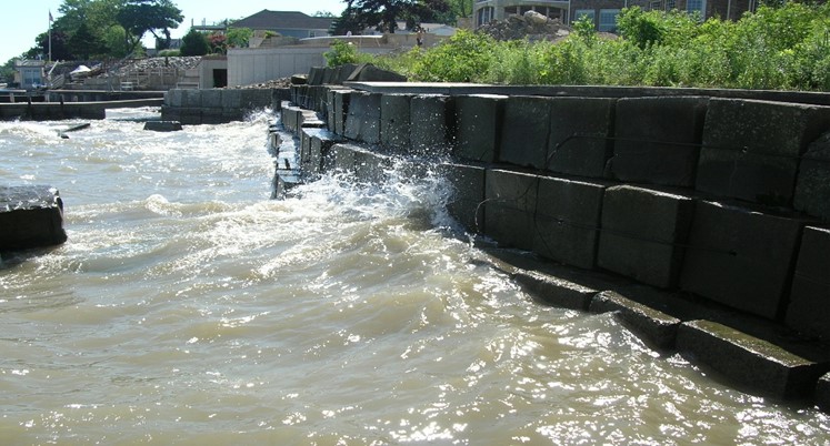
According to the United Nations, 40% of the world’s population lives within 100km of a coastline and this number is projected to increase. Coastal protection structures, typically made of rock, steel and concrete, are used to protect homes and businesses from waves, storm surges and flooding. On Lake Erie’s shoreline in Ohio, 80% of the shoreline is protected with these simple and rigid materials.
Example of revetment – provided by ODNR
Example of seawall provided by ODNR
Worldwide, shoreline hardening destroys the land-water interface and nearshore habitat complexity, key to many significant transitional ecosystems and nursery habitat for fish, birds and other species. These natural coastal ecosystems also often act as protective barrier from waves, storm surges and flooding.
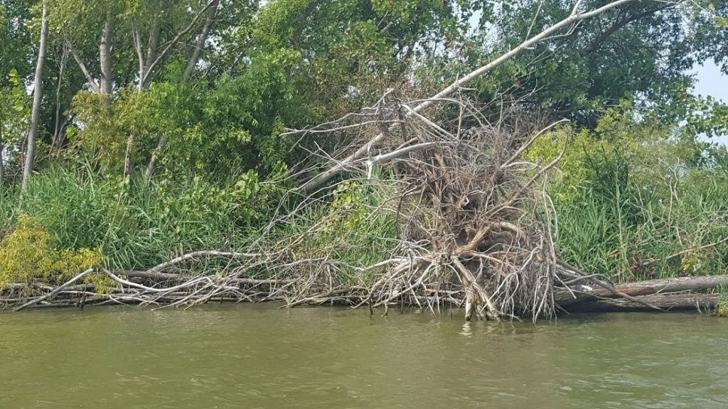
Natural shoreline example – Downed tree with root overhang in Sandusky Bay – August 2018
Check out some examples of creative infrastructure and restoration efforts in marine environments for more inspiration: ECOncrete, Reef Design Lab, TetraPOT and Cemex.
Click here for more information on Dr. Gruber’s lab.
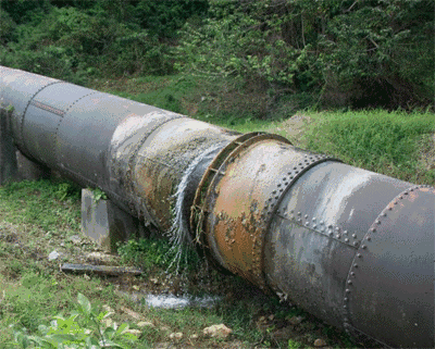
Figure 1: The aftermath of microorganisms accelerating pipeline corrosion
Figure 2: Sulfate reducing bacteria are good examples.
Background:
Microbially Induced Corrosion (MIC) is the deterioration of metals that is brought about by the metabolic activity of various microorganisms. The phenomenon is very detrimental to infrastructure and can accelerate the rates at which metals degrade, resulting is costly repairs to the affected areas. A major problem of MIC is the fact that it can be very difficult to monitor in some places, such as pipelines and sewer lines. While biocides may be used in order to kill the bacteria that have formed on the metals, the solutions are not always environmental friendly and can also be extremely exorbitant.
Current Research Projects:
Presently, we are trying to find more effective ways to monitor the effects of MIC through the use of electrochemical techniques to monitor current flow, electron acceptance and electron donation between metals and microorganisms. The goal is to preemptively catch problems in a pipe system before irreparable damage has been done. Performing chemical analyses on water samples is also crucial, as it helps guide necessary modifications for techniques based on the findings.
Click here to learn more about Dr. Senkos’ lab.
Figure 1: Andronowski Lab ‘Beam Team’ members during experiments at the Canadian Light Source national synchrotron facility in Saskatchewan, Canada. From left to right: Former UA undergraduate Linda Muakkassa, UA doctoral student Reed Davis, Dr. Andronowski, and former UA undergraduate Hannah Stephen.
What do forensic science, bones, X-ray beams, and 3D imaging have in common? The Andronowski Lab!
Research background:
The estimation of the postmortem interval (PMI), or time since death, is a crucial and fundamental consideration in forensic death scene investigations. With the progression of time and soft tissue decomposition, a postmortem estimation by a medical examiner becomes more difficult and less accurate, as bone and teeth are often the only remaining tissues of an organism. Diagenetic agents (e.g., microbial infiltration, ground water, soil, pH balance, fungi) are known to structurally alter bone tissue micromorphology over time. This may further disrupt high-quality DNA samples or histological age-at-death estimates in forensic anthropological contexts. To date, there are no quantitative tools available to the forensic anthropologist to evaluate bone diagenesis.
Figure 2: Current Tiered Mentoring student, Gina Tubo, during experiments at the Canadian Light Source.Project details:
Goal: To identify and quantify bone tissue microarchitectural changes across increasing postmortem intervals and evaluate how various diagenetic agents impact microstructures of cortical bone used in histological age estimation methods in forensic anthropology.
Methods: An innovative 3D X-ray imaging technique (Synchrotron Radiation-based micro-CT) will be utilized. High-resolution imaging experiments will be executed at the Canadian Light Source national synchrotron facility.
Sample groups: Four predetermined postmortem intervals (0-3 years, 4-10 years, 11-20 years and >20 years) were identified and three skeletons selected to represent each interval (n=12).
Figure 3: Dr. Andronowski visualizing cortical bone data at the Canadian Light Source.Benefits for the Student:
Learn about how we apply the principles of bone remodeling to estimate age-at-death in forensic anthropology
Receive training in 3D X-ray imaging using desktop micro-CT, big data processing, the preparation of bone tissue slides, and dissection techniques.
As a Tiered Mentoring student, you may have the opportunity to join the Andronowski Lab ‘Beam Team’ and assist with imaging experiments for this project.
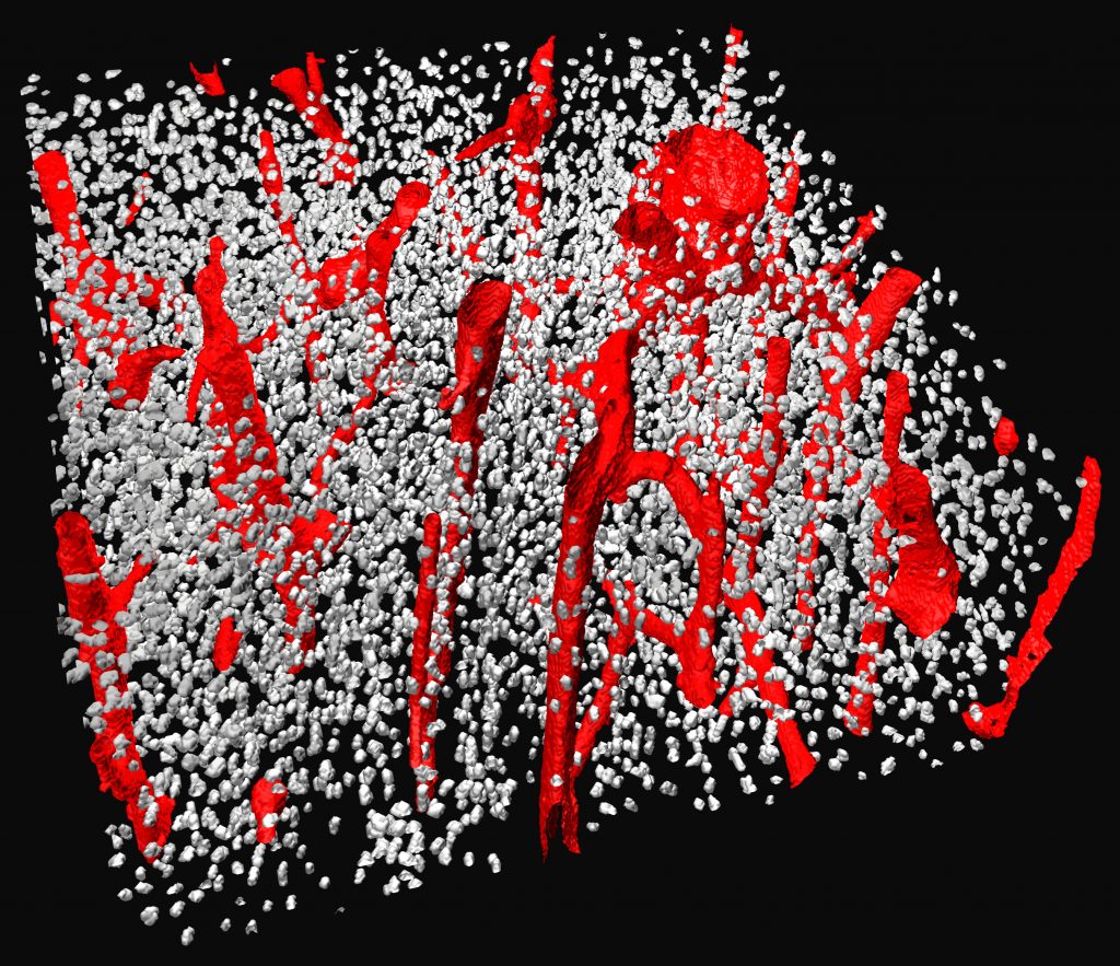
Figure 4: 3D render of cortical bone showing vascular porosity (red) and bone cellular spaces (grey). Data collected at the Canadian Light Source.Qualifications:
Dr. Andronowski is interested in undergraduate students from any major who are willing to learn the above skills. Preference will be given, however, to those who have experience with dissection (animal and/or human), histological techniques, and computer programming/modeling.
Click here to learn more about Dr. Andronowski’s lab
Figure 2. Shewanella oneidensis MR-1 growing on hematite, using iron as an electron acceptor.
link: https://www.livescience.com/28163-bio-batteries-one-step-closer.htmlBackground:
Banded iron formations (BIF) are the world’s largest and most widespread source of iron. The Carajas BIF of Brazil is associated with the presence of a high-grade iron ore and extensive cave development. The formation of these caves appears to be through microbial iron reduction, driven by the organic carbon from high surficial primary productivity in overlying soils. In order for the ore to form, it must be depleted of silica, yet there is no clear explanation of how the silica is being mobilized. Silica has a low solubility in water and is difficult to dissolve without a catalyst at circumneutral pH. We know iron reduction is occurring within the caves and this changes iron to its soluble form, but there is no redox reaction for silica. This suggests the possibility that iron redox reactions influence the dissolution of silica.
Current Research Projects:
We are trying to understand the process of silica mobilization through microbially driven iron redox reactions, using bacterial cultures grown in both anaerobic and aerobic conditions with iron and silica.
To analyze these bacterial cultures, we will do both geochemical and biological analyses.
Skills you will develop:
Culturing bacteria · Growing bacteria in anaerobic conditions and using an anaerobic chamber · Creating various forms of media · Various geochemical analyses · Using a centrifuge, autoclave, pH meter, pipettes, spectrometer, ion chromatographer, microscope · You will begin to gain an understanding of the complex relationships between microbial life and geological processes.
Click here to learn more about Dr. Senkos’ lab.