Evaporative systems inspired by leaf designs

[Past Projects]

Dr. Petra Gruber and Ariana Rupp


In our biomimicry design lab, we are interested in learning from leaf shapes. Leaves not only are plants’ solar cells, they also pull nutritious water through plant tissues via transpiration.

Because of this, leaves continuously endure energy and water flows, with leaf structure shaping such flows, potentially helping dissipation of water vapor and excessive heat.

Can we discover structural lessons in leaf design, to apply to human technologies which also uses evaporation for thermal management?


There are two sides to learning from leaf thermodynamics and shapes:

  • Biology efforts involve studying leaf exchange from real plants under different environmental conditions and collecting data related to leaf shape, water status and temperature.
  • The engineering approach deals with application of design lessons, testing of leaf-inspired models in controlled environments, precise measuring and data analysis.


FOR MORE INFO, PLEASE CONTACT:

pgruber@uakron.edu

ak230@zips.uakron.edu


Click here for more information on Dr. Gruber’s lab.

Protect our coasts – designing lab-scale wave simulation studies of Lake Erie

[Past Projects]

Dr. Teresa Cutright, Dr. Henry Astley and Elena Stachew


According to the United Nations, 40% of the world’s population lives within 100km of a coastline and this number is projected to increase. Coastal protection structures, typically made of rock, steel and concrete, are used to protect homes and businesses from waves, storm surges and flooding. On the Lake Erie shoreline in Ohio, 80% of the shoreline is hardened/protected with concrete, steel and rock.


Example of revetment – provided by Jim Park, ODNR                      Example of seawall – provided by Jim Park, ODNR

Lab-scale hydrodynamic studies will be conducted using a 11 ft. re-circulating flume in the Hydraulics Lab of the Civil Engineering Department.  The studies will test next-generation biomimetic forms and biologically friendly materials for coastal protection structures that are more compatible with aquatic life in Lake Erie.

You will help build a wave plate and actuator system using Arduino components to create realistic lake wave profiles in the flume. You will also help build a program in Python for real-time video tracking of changing wave dynamics. There may be an opportunity to build smaller-scale wave tanks for use in classrooms and other public educational settings. Check out this Youtube video for an idea!


Recirculating flume at University of Akron’s Hydraulics Lab

It is highly recommended (though not required) that you pair this tiered mentoring project with enrollment in the Special Topics undergraduate biology course 3100-495:007 Digital Skills for Biologists (3 credits). Expect to learn about 3D modeling, 3D printing, videography and image tracking, in addition to Lake Erie’s natural history, food web dynamics and systems ecology. We are interested in undergraduate students from any major that are willing to learn any of the above skills and subject areas. No prior experience is necessary.

Work at the interface of engineering and biology to improve nearshore aquatic habitat conditions on Lake Erie with a Biomimicry Fellow.


Click here to learn more about Dr. Cutright’s lab

Click here to learn more about the Astley Lab

Long-term Effects of Prolonged Opioid Use on Cortical Bone Remodeling

[Past Projects]

Dr. Janna Andronowski and Reed Davis


The misuse and addiction to opioids (and synthetic opioids) is a serious public health crisis nationwide that has become an epidemic. Current evidence suggests that opioids upset the balance of bone remodeling towards more destruction and less formation of bone. Experimental studies have been limited by the fact that small laboratory animals traditionally used in bone research (mice and rats) do not exhibit spontaneous cortical bone remodeling, making them a poor choice of animal model for this subject.

A new project in the Andronowski Lab seeks to develop a long-term model for studying the effects of prolonged opioid use on cortical bone remodeling in an animal which remodels its cortical bone in a manner comparable to humans, the rabbit. An innovative 3D X-ray imaging technique (micro-CT), combined with dynamic histomorphometry, will allow us to describe how morphine and fentanyl affect microscopic structures of cortical bone used in histological age estimation methods in forensic anthropology. Given the limited data available related to the impact of opioid abuse on bone remodeling, our goal is to further understandings of the underlying biological processes and improve the applicability of histological age-estimation methods and scientific standards within the field of forensic anthropology.

Prospective students can expect to learn about how we apply the principles of bone remodeling to estimate age-at-death in forensic anthropology, 3D X-ray imaging using desktop micro-CT (Figure 3), big data processing, the preparation of bone tissue slides, animal handling, and dissection techniques. Dr. Andronowski is interested in undergraduate students from any major who are willing to learn the above skills. Preference will be given, however, to those who have experience with live animal research, animal dissections, histological techniques, and computer programming/modeling.


Figure 1: Coupled osteoclast and osteoblast activity depicted as a Basic Multicellular Unit during bone remodeling. A) Osteoclastic activity near the leading edge of the cutting cone, B) initiation of osteoblastic activity, C) active osteoblast activity, and D) a fully formed intact osteon depicting a Haversian Canal in the center.


Figure 2: Micro-CT 3D render of cortical bone showing vascular porosity (red) and osteocyte lacunae (grey). Data collected at the Canadian Light Source synchrotron.


Figure 3: SkyScan 1172 Desktop Micro-CT system at the University of Akron.


Click here to learn more about Dr. Andronowski’s lab

Mycelium material systems

[Past Projects]

Dr. Petra Gruber and Thibaut Houette


This project is about the use of fungi to solidify agricultural waste aggregate to solid materials that can be used for construction. The goal is to examine the currently used growth processes, and find out about improvements and practical solutions for small scale production, to produce materials and do a variety of tests on the properties. The research will be done in collaboration with phD student Thibaut Houette, and take place in the Biology labs. The student should be interested in fungi microbiology and their cultivation, and open to an interdisciplinary approach to develop biomimetic solutions.


Click here for more information on Dr. Gruber’s lab.

Using Robots to Understand Fish Maneuverability

[Past Projects]

Dr. Henry Astley and Stephen Howe


Fish are adept swimmers that control their locomotion using relatively simple methods. We have been gathering swimming data from live fish and creating models to describe their behaviors. Working with live fish comes with certain limitations due to unpredictable fish behavior and the inaccessibility of rare or delicate species. We are developing a robotic model to test hypotheses about turning behavior in fish that we would not be able to test with live fish. This robot will help us understand how fish control turns and what are the limitations of their maneuverability. The robot will allow us to study the relationship between body shape and swimming performance to add some perspective to a long-standing debate in the fish swimming literature. Expect to learn about robotics, 3D modeling, videography and image tracking, and fish biomechanics. We are interested in undergraduate students from any major that are willing to learn any of the above skills. No prior experience is necessary.


Click here to learn more about the Astley Lab


Sticky When Wet: Understanding Tree Frog Adhesion on Different Surfaces

[Past Projects]

Dr. Henry Astley, Dr. Peter Niewiarowski, Derek Jurestovsky, and Austin Garner


Research Description

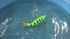
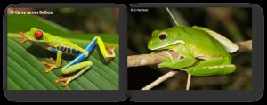
Tree frogs secrete a mucous on the bottom of their micropatterned toe pads that permit them to adhere to surfaces like two wet glass microscope slides stuck together (capillary adhesion). Theoretically, tree frog adhesion should not be dependent on whether the surface of interest repels or attracts water because the secreted mucous is capable of sustaining adhesive contact on a variety of surfaces. In this study, we will measure live tree frog adhesion under dry, misted with water, and wet conditions using a variety of artificial surfaces that range in their water repellency.

Benefits of this Research Experience

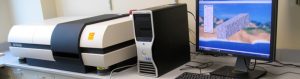
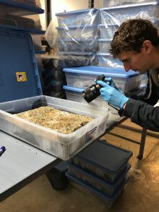
Undergraduate students in the Astley and Niewiarowski labs will gain an array of critical research skills including: amphibian care and handling, live animal performance measurements, experimental design, statistics, microscopy, surface characterization methods, scientific writing, and more. Additionally, many of the undergraduate students in our labs have been co-authors on several papers published in peer-reviewed journals (see below)!


*Denotes undergraduate student

Niewiarowski, P. H., Lopez, S., Ge, L., Hagan, E.* and Dhinojwala, A. (2008). Sticky gecko feet: the role of temperature and humidity. PLoS. ONE 3, e2192.

Niewiarowski, P. H., Stark, A., McClung, B.*, Chambers, B.* and Sullivan, T.* (2012). Faster but Not Stickier: Invasive House Geckos Can Out-Sprint Resident Mournful Geckos in Moorea, French Polynesia. J. Herpetol. 46, 194-197.

Stark, A. Y., Sullivan, T. W.* and Niewiarowski, P. H. (2012). The effect of surface water and wetting on gecko adhesion. J. Exp. Biol. 215, 3080-6.

Stark, A. Y., Badge, I., Wucinich, N. A.*, Sullivan, T. W.*, Niewiarowski, P. H. and Dhinojwala, A. (2013). Surface wettability plays a significant role in gecko adhesion underwater. Proc Natl Acad Sci U S A 110, 6340-5.

Stark, A. Y., Wucinich, N. A.*, Paoloni, E. L.*, Niewiarowski, P. H. and Dhinojwala, A. (2014). Self-drying: a gecko’s innate ability to remove water from wet toe pads. PLoS ONE 9, e101885.

Stark, A. Y., McClung, B.*, Niewiarowski, P. H. and Dhinojwala, A. (2014). Reduction of water surface tension significantly impacts gecko adhesion underwater. Integr. Comp. Biol 54, 1026-33.

Badge, I., Stark, A. Y., Paoloni, E. L.*, Niewiarowski, P. H. and Dhinojwala, A. (2014). The role of surface chemistry in adhesion and wetting of gecko toe pads. Scientific Reports 4, 6643.

Stark, A. Y., Ohlemacher, J.*, Knight, A.* and Niewiarowski, P. H. (2015). Run don’t walk: locomotor performance of geckos on wet substrates. J. Exp. Biol. 218, 2435-41.

Stark, A. Y., Palecek, A. M.*, Argenbright, C. W., Bernard, C.*, Brennan, A. B., Niewiarowski, P. H. and Dhinojwala, A. (2015). Gecko adhesion on wet and dry patterned substrates. PLoS. ONE 10, e0145756.

Stark, A. Y., Dryden, D. M., Olderman, J.*, Peterson, K. A., Niewiarowski, P. H., French, R. H. and Dhinojwala, A. (2015). Adhesive interactions of geckos with wet and dry fluoropolymer substrates. J. R. Soc. Interface 12, 20150464.

Stark, A. Y., Subarajan, S.*, Jain, D., Niewiarowski, P. H. and Dhinojwala, A. (2016). Superhydrophobicity of the gecko toe pad: biological optimization versus laboratory maximization. Phil. Trans. R. Soc. A 374, 20160184.

Klittich, M. R., Wilson, M. C., Bernard, C.*, Rodrigo, R. M.*, Keith, A. J.*, Niewiarowski, P. H. and Dhinojwala, A. (2017). Influence of substrate modulus on gecko adhesion. Scientific Reports 7, 43647.

Garner, A. M.*, Stark, A. Y., Thomas, S. A. and Niewiarowski, P. H. (2017). Geckos go the Distance: Water’s Effect on the Speed of Adhesive Locomotion in Geckos. J. Herpetol. 51, 240-244.


Click here to learn more about the Astley Lab

Click here to learn more about the Niewiarowski lab

Investigating the morphology of melanopsin ganglion cells in health and disease

[Past Projects]

Dr. Jordan Renna and Katelyn Sondereker


In general, our lab studies the eye: We study the retina and how it develops. More specifically, we are interested in the development of melanopsin ganglion cells. Melanopsin ganglion cells are like rods and cones, but they send the brain non-visual information about circadian rhythms and the pupillary light reflex. Melanopsin ganglion cells are also a target for studies involving health and disease, since they are resistant to many visual disorders.

My current project involves studying the morphology of melanopsin ganglion cells in a mouse model of glaucoma. Staining retinal tissue to visualize melanopsin will allow us to determine how the anatomy of melanopsin ganglion cells changes as the disease progresses. This will provide invaluable information about the effect of glaucoma on melanopsin ganglion cells. I am looking for students to become independent in their ability to perform eye dissections (Fig. 1, 2), stain tissue with immunohistochemistry (Fig. 3), and trace the morphology of melanopsin ganglion cells with programs like ImageJ (Fig. 4). These techniques will be useful to anyone interested in a career in the medical field or in biomedical and clinical research.


Figure 1: A lateral view of an enucleated mouse eye before dissection.


Figure 2: An isolated mouse retina immunostained for s-opsin (blue).


Figure 3: Retinal tissue immunostained for melanopsin (magenta) and VGLUT2 (green).


Figure 4: A whole-cell reconstruction tracing of a melanopsin ganglion cell.


Click here for more information on Dr.Renna’s lab.

Macro-invertebrates 3D modeling

[Past Projects]

Dr. Francisco Moore and Banafsheh Khakipoor


Do you aspire to come up with creative ways to teach youth? Are you excited about 3D printers? If yes, then consider applying for this project.

This project is a collaboration with Cleveland Metro Parks to develop a K-12 curriculum for water quality monitoring by learning about macro-invertebrates. We create three-dimensional models of the critters living in a watershed to use as a teaching tool. This is especially beneficial when students cannot go to a water body to study these invertebrates in their habitats due to weather, accessibility, etc. An indoor classroom can mimic a river outline and use the 3D models as replicas.

In this project we will use and learn:

  • Collect Macro-invertebrates from Watershed Stewardship Center
  • Use a Micro CT scanner to scan these marco-invertebrates.
  • Use ImageJ (image analysis software) to create 3D models using series of images taken by MicroCT scanner.
  • Print the models using a 3D printer.

Click here for more information on Dr. Moore’s lab.

Tracking human health & diet with stable isotopes

[Past Projects]

Dr. Anne Wiley and Nate Michael


Could you tell what a stranger ate during the past month, using only a tiny sample of their hair? Could you predict their risk of diabetes, hypertension, or other chronic disease? These are the questions at the heart of our research… and potentially at the heart of your new project.

Unbeknownst to most people, our dietary history is recorded by subtle shifts in the chemical composition of our hair. Stable isotopes are alternative forms of an element that differ in their number of neutrons (e.g. 14N has 7 neutrons while 15N has 8); the ratio of these isotopes in human tissues are inherited from our food. For example, people who consume meat and other animal-based foods have more 15N in their hair than vegans. And the more corn syrup people consume, the more 13C will become concentrated in their blood. Because stable isotopes reflect aspects of diet such as sugar consumption that are linked with obesity and chronic disease, many researchers have predicted that they will have a powerful impact in future human health studies.

The goal of this project is to collect hair samples from University of Akron students and determine exactly what aspects of diet can (and can’t) be quantified using stable isotope techniques.  We’re searching for a dedicated undergraduate who can help to collect hair samples from volunteers, help administer diet and health surveys, learn to analyze samples for their isotopic content in lab, and push our understanding about the isotope-diet link to the next level. The recruited student will join an interdisciplinary team (ecologists and stable isotope specialists, an anthropologist, and an expert in human movement) who are aiming to apply stable isotopes to the study of food availability and health in low-income Cleveland neighborhoods. The student may have the opportunity to join in the Cleveland-based research, depending on their timeline.


Click here to learn more about Dr. Wiley’s lab.

Classifying Ion Channels in the Retina

[Past Projects]

Dr. Jordan Renna and Matthew Tarchick


Our lab studies the development of circuits in the retina. During development, an interconnected layer of the retina called the starburst amacrine cell layer fires bursting patterns. These patterns occur for 10-12 days. A specific interest to me is the how potassium and calcium conductance plays a modulating role in these bursting patterns, and how those bursts can pass on information. To better understand conductance, we are exploring the expression of ion channel proteins and genes, as well as visualizing their location, and using agents which inhibit those channel activities.

To study this, we use many typical molecular biology methods like immunohistochemistry, western blotting and quantitative PCR. With western blotting we can get an idea of the change in protein expression and monitor the presence of a protein in the retina. With Immunohistochemistry we can get a better look at the localization of a certain protein. I am looking for students that can help me by learning these techniques. These techniques are very valuable for careers in medical research and various other biomedical studies.


Fig 1. Retinal Lysate Western Blot of Beta Actin


Fig 2. Immunohistochemistry:

Blue is DAPI, a nuclear stain

Red is choline acetyltransferase

Green is small conductance potassium channel 1


Click here for more information on Dr.Renna’s lab.