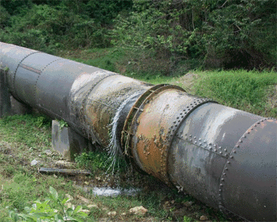
Figure 1: The aftermath of microorganisms accelerating pipeline corrosion
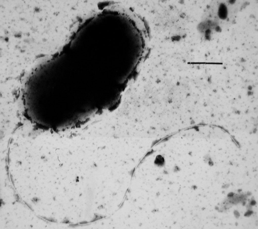
Figure 2: Sulfate reducing bacteria are good examples.
Background:
Microbially Induced Corrosion (MIC) is the deterioration of metals that is brought about by the metabolic activity of various microorganisms. The phenomenon is very detrimental to infrastructure and can accelerate the rates at which metals degrade, resulting is costly repairs to the affected areas. A major problem of MIC is the fact that it can be very difficult to monitor in some places, such as pipelines and sewer lines. While biocides may be used in order to kill the bacteria that have formed on the metals, the solutions are not always environmental friendly and can also be extremely exorbitant.
Current Research Projects:
Presently, we are trying to find more effective ways to monitor the effects of MIC through the use of electrochemical techniques to monitor current flow, electron acceptance and electron donation between metals and microorganisms. The goal is to preemptively catch problems in a pipe system before irreparable damage has been done. Performing chemical analyses on water samples is also crucial, as it helps guide necessary modifications for techniques based on the findings.
Click here to learn more about Dr. Senkos’ lab.
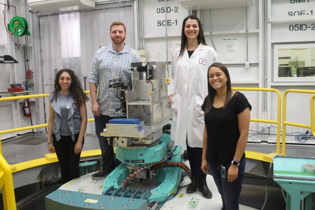
Figure 1: Andronowski Lab ‘Beam Team’ members during experiments at the Canadian Light Source national synchrotron facility in Saskatchewan, Canada. From left to right: Former UA undergraduate Linda Muakkassa, UA doctoral student Reed Davis, Dr. Andronowski, and former UA undergraduate Hannah Stephen.
What do forensic science, bones, X-ray beams, and 3D imaging have in common? The Andronowski Lab!
Research background:
The estimation of the postmortem interval (PMI), or time since death, is a crucial and fundamental consideration in forensic death scene investigations. With the progression of time and soft tissue decomposition, a postmortem estimation by a medical examiner becomes more difficult and less accurate, as bone and teeth are often the only remaining tissues of an organism. Diagenetic agents (e.g., microbial infiltration, ground water, soil, pH balance, fungi) are known to structurally alter bone tissue micromorphology over time. This may further disrupt high-quality DNA samples or histological age-at-death estimates in forensic anthropological contexts. To date, there are no quantitative tools available to the forensic anthropologist to evaluate bone diagenesis.
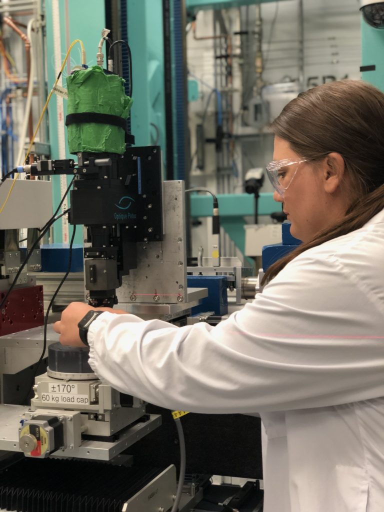
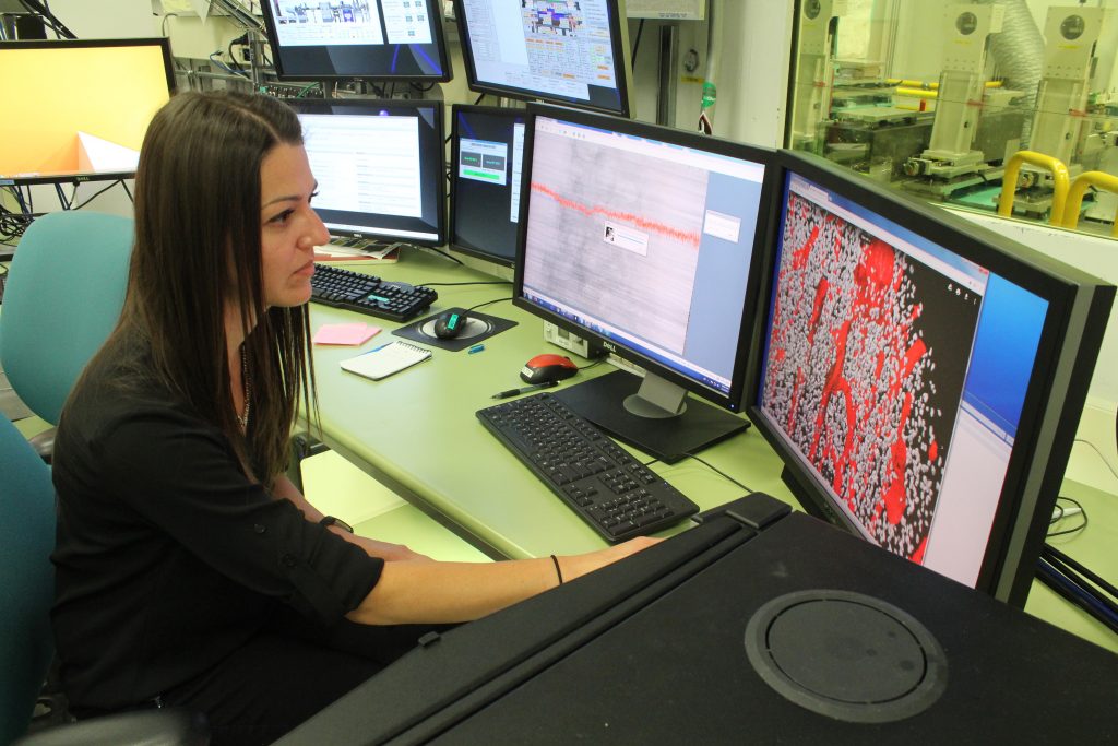
Figure 2: Current Tiered Mentoring student, Gina Tubo, during experiments at the Canadian Light Source.Project details:
Goal: To identify and quantify bone tissue microarchitectural changes across increasing postmortem intervals and evaluate how various diagenetic agents impact microstructures of cortical bone used in histological age estimation methods in forensic anthropology.
Methods: An innovative 3D X-ray imaging technique (Synchrotron Radiation-based micro-CT) will be utilized. High-resolution imaging experiments will be executed at the Canadian Light Source national synchrotron facility.
Sample groups: Four predetermined postmortem intervals (0-3 years, 4-10 years, 11-20 years and >20 years) were identified and three skeletons selected to represent each interval (n=12).
Figure 3: Dr. Andronowski visualizing cortical bone data at the Canadian Light Source.Benefits for the Student:
Learn about how we apply the principles of bone remodeling to estimate age-at-death in forensic anthropology
Receive training in 3D X-ray imaging using desktop micro-CT, big data processing, the preparation of bone tissue slides, and dissection techniques.
As a Tiered Mentoring student, you may have the opportunity to join the Andronowski Lab ‘Beam Team’ and assist with imaging experiments for this project.
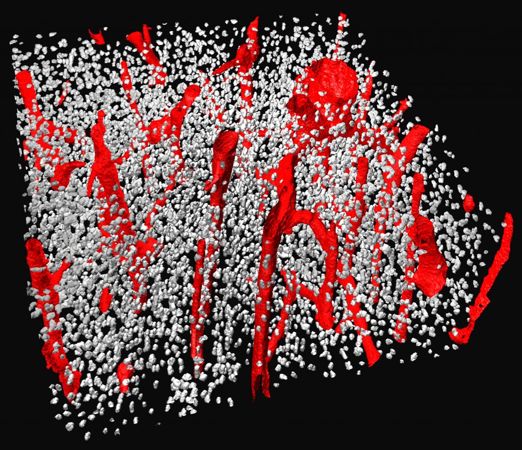
Figure 4: 3D render of cortical bone showing vascular porosity (red) and bone cellular spaces (grey). Data collected at the Canadian Light Source.Qualifications:
Dr. Andronowski is interested in undergraduate students from any major who are willing to learn the above skills. Preference will be given, however, to those who have experience with dissection (animal and/or human), histological techniques, and computer programming/modeling.
Click here to learn more about Dr. Andronowski’s lab
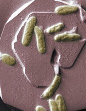
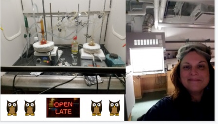
Figure 2. Shewanella oneidensis MR-1 growing on hematite, using iron as an electron acceptor.
link: https://www.livescience.com/28163-bio-batteries-one-step-closer.htmlBackground:
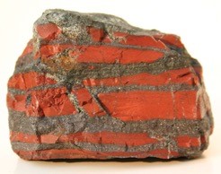
Banded iron formations (BIF) are the world’s largest and most widespread source of iron. The Carajas BIF of Brazil is associated with the presence of a high-grade iron ore and extensive cave development. The formation of these caves appears to be through microbial iron reduction, driven by the organic carbon from high surficial primary productivity in overlying soils. In order for the ore to form, it must be depleted of silica, yet there is no clear explanation of how the silica is being mobilized. Silica has a low solubility in water and is difficult to dissolve without a catalyst at circumneutral pH. We know iron reduction is occurring within the caves and this changes iron to its soluble form, but there is no redox reaction for silica. This suggests the possibility that iron redox reactions influence the dissolution of silica.
Current Research Projects:
We are trying to understand the process of silica mobilization through microbially driven iron redox reactions, using bacterial cultures grown in both anaerobic and aerobic conditions with iron and silica.
To analyze these bacterial cultures, we will do both geochemical and biological analyses.
Skills you will develop:
Culturing bacteria · Growing bacteria in anaerobic conditions and using an anaerobic chamber · Creating various forms of media · Various geochemical analyses · Using a centrifuge, autoclave, pH meter, pipettes, spectrometer, ion chromatographer, microscope · You will begin to gain an understanding of the complex relationships between microbial life and geological processes.
Click here to learn more about Dr. Senkos’ lab.
Would you like an opportunity to be part of a lab that plans to SAVE THE WORLD?! Then our interdisciplinary lab is the place for you! Currently, we have eight undergraduate students working on various degrees (biology, geology, chemistry, and biomechanics). We all support each other in EVERYTHING we do. We would love to add YOU to our crew! *I support independent undergraduate research ideas within the scopes of our lab!*
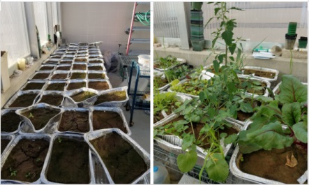
Phytoremediation of lead-contaminated soils: Approximately 400 plants grew in our greenhouse in high levels of lead (plus the control, lead-free)! We chemically isolate the lead from the plant tissues and analyze by ICP (Inductively Coupled Plasma). To date, we have found several native species and a food species having high concentrations of lead in the tissues! Needs for this project: digesting and refluxing plant tissues, lab clean-up, data entry, analyzing data, and writing reports.Potential future projects: grind, digest, isolate lead from root tissues of plants, data entry, analyze data and write reports.
Plants on Mars/Super Powers of Plants Part 2: I want to play with the idea of plants surviving on Mars. Plants survive in extreme environments on Earth and often multiple environmental stressors work to increase survival against the odds. This fascinating fact about plants suggests the possibility of plants tolerating certain environmental conditions on Mars. We will grow plants in NASA-approved regolith and subject them to several Martian conditions, assessing the impacts on plant growth! Ideally, all undergraduates working on the Mars project will participate in BURS Biology Undergraduate Research Symposium before they graduate (held in spring semesters). Needs for this project: reliability and dedication to care for plants, thorough data keeping, accurate measuring, data entry and analysis.Lab clean-up is an ongoing duty and part of all research.
Conservation Education at Cleveland Metroparks Zoo: My proposed research focuses on understanding individuals who actively participate in advocating for wildlife conservation at Cleveland Metroparks Zoo, where I am a Biomimicry Education Fellow. Specifically, I will investigate higher-level advocates: Zoo Crew (teen volunteer program), Young Professionals (20-40 year-old professionals who advocate at CMZ), and Advanced Inquiry Program (Master’s degree students from Miami University) through surveys, interviews, social media analytics, and behavioral observations. Needs for this project: survey development, interview transcription/coding/categorization (TBD), social media tracking (i.e. frequency of specified types of shared posts during a specified time period), behavioral observations in person (i.e. making notes of personal interactions, physical actions, etc.), data entry, data coding, data analysis, report writing, and potentially more!
Biomimicry Education and Outreach: As a Biomimicry Fellow, I do a lot of public outreach in Northeast Ohio (zoos, museums, public and private K-12 schools and universities, Women in Science events, NASA-Glenn, non-profit art groups, community centers, etc.). Our lab students are always welcome to join!
Words from current students:
“Having the opportunity to participate in the tiered mentoring program has exposed me to an extensive amount of lab experience that I otherwise would not have had, improving my capabilities in a lab environment on both a conceptual and practical level. The program has also provided an excellent collaborative atmosphere in which I am able to make meaningful contributions to ongoing research projects and receive valuable guidance and support on any projects I choose to undertake on my own.” Gavin DeMali, 2nd year Tiered Mentoring student “I would like to start out and say that I have had a great time in the tiered mentoring program. Everyone was so welcoming and willing to help me out, which means a lot to me. Rebecca never has a problem assisting me with information on the lab or any questions in general. Her bubbly personality makes me feel at home and in a safe environment. Each person I have met in the lab has unique attributes that make working there all the more enjoyable. Not only do the people make the experience rewarding, but the lab’s organization as well. Whenever I have ran samples late I end up doing my homework in between, so it creates an atmosphere of productivity. Not to mention, I appreciate the diversity in plants that are tested. Also I love how we are able to take some plants home! One thing I would change about my time with the tiered mentoring program is the time I spent. I would like to dedicate more time in the lab and eventually come up with a research proposal that I can conduct myself! Thank you for this cool opportunity!” Olivia Orr, 1st year Tiered Mentoring student
“I believe the tiered mentoring program is a great asset to the university and every research lab. Throughout my past years of doing research, I have gained multiple skills that will benefit me in a future job or in life in general. One of the main reasons why I got my environmental science internship was that I had experience working independently and also with a team within our research lab. I was able to tell my employers that I had experience working with chemical equipment which helped me in the field as well. It has not only helped to open doors for my future career but has also further my education. I am very fortunate to have the opportunity to work in our research lab and I highly recommend anyone who is interested to explore their options.” – Stephanie Sawicki
Click here for other information about Dr. Mitchell’s lab.
Dr. Teresa Cutright, Dr. Henry Astley and Elena Stachew
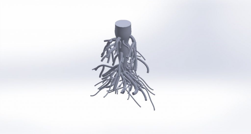
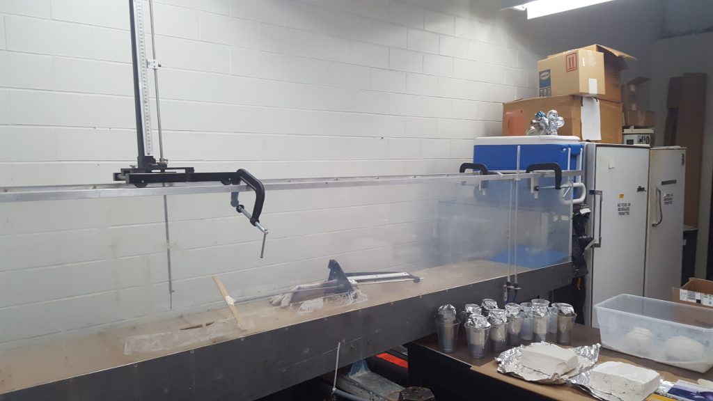
We are studying the ability to quickly design, prototype and evaluate biomimetic forms to restore natural habitat complexity to our lake. Lab-scale wave attenuation and sediment depositional prediction studies are conducted using a 11 ft. re-circulating flume in the Hydraulics Lab of the Civil Engineering Department. We are primarily testing complex root forms mimicked after native coastal forests along Lake Erie. Other root forms may be considered and explored, such as those mimicked after mangroves or coastal wetland plants.
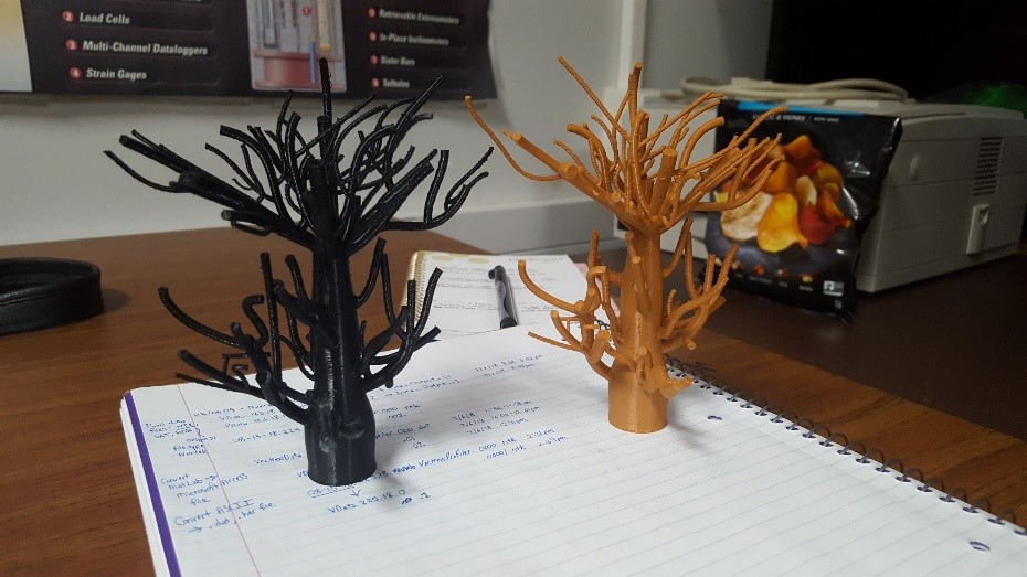
3D printed PLA root structures from UA MakerspaceWhite oak Solidworks root model – from Liang T, Knappett JA, et al. 2017 paper in Landslides
You’ll gain skills in one or more of the following:
Simple electronics
Programming in Python
3D modeling and printing
Field visits to Lake Erie and surrounding watersheds
Materials investigation
There may be an opportunity to build smaller-scale wave tanks for use in classrooms and other public educational settings. Check out this Youtube video for an idea!
Wave tank at University of Akron’s Hydraulics Lab
We are interested in undergraduate students from any major that are willing to learn and explore. No prior experience is necessary. Work at the interface of engineering and biology to improve the coastal ecology of Lake Erie with a Biomimicry Fellow.
Additional Background:
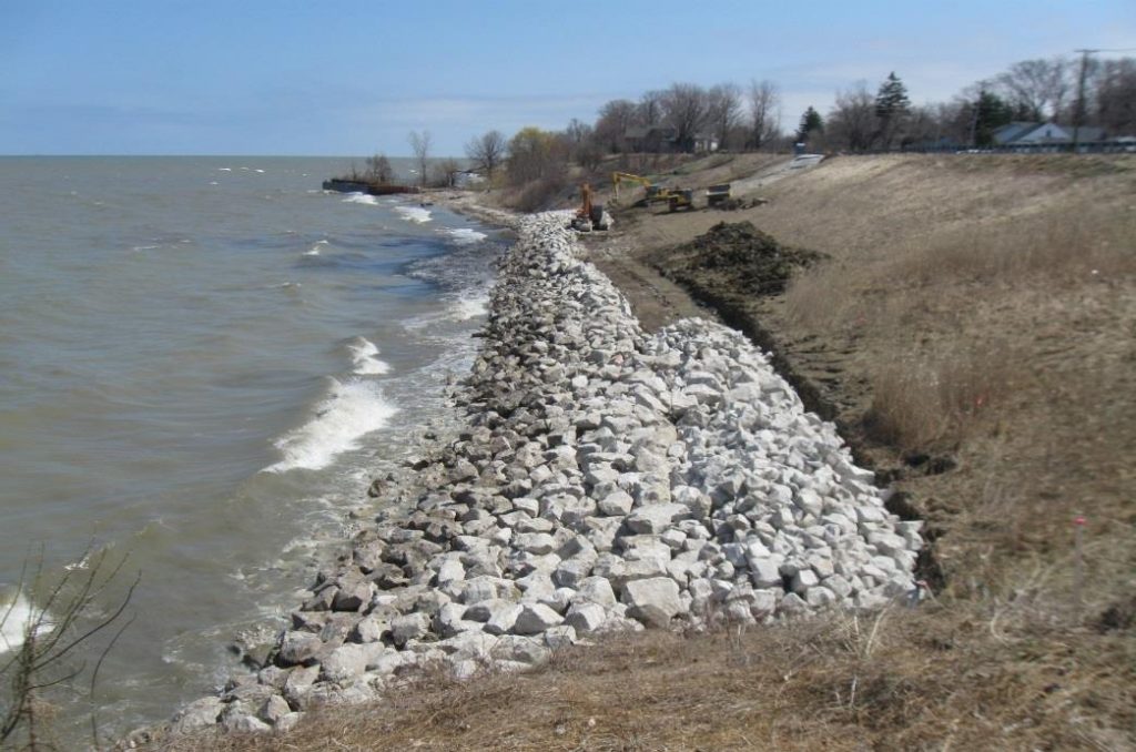
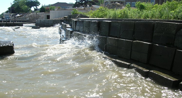
According to the United Nations, 40% of the world’s population lives within 100km of a coastline and this number is projected to increase. Coastal protection structures, typically made of rock, steel and concrete, are used to protect homes and businesses from waves, storm surges and flooding. On Lake Erie’s shoreline in Ohio, 80% of the shoreline is protected with these simple and rigid materials.
Example of revetment – provided by ODNRExample of seawall – provided by ODNR
Worldwide, shoreline hardening destroys the land-water interface and nearshore habitat complexity, key to many significant transitional ecosystems and nursery habitat for fish, birds and other species. These coastal ecosystems also often act as natural protection from waves, storm surges and flooding.
Natural shoreline example: Downed tree with root overhang in Sandusky Bay (August 2018)
Check out some examples of creative infrastructure and restoration efforts in marine environments for more inspiration: ECOncrete, Reef Design Lab, TetraPOT and Cemex.
Click here to learn more about Dr. Cutright’s labClick here to learn more about the Astley Lab
Dr. Todd Blackledge, Dr. Ali Dhinojwala, Angela Alicea-Serrano and K Zin Htut
Buehler et al. NanoWerk. 2010; Zhang and Tso et al. Extracellular Composite Matrices in Arthropods, 2016, Springer, First Edition; Angnarrson et al. Plos One. 2010, 5, 9, 1-8.
Research Description
Spider dragline silk is the toughest biomaterial in nature; spider silk is ounce for ounce tougher than steel and the Kevalr in bullet resistant armor. Silk properties and composition inspire genetically engineered biomaterialssuch as medical sutures, artificial tendons and ligaments, which must be extensible and strong but also light. These future applications of spider silk require a clear understanding of protein structure and its connection with the mechanical properties of biomaterials.
We are investigating the world’s toughest dragline silk from Darwin’s bark spider, which spins giant orb webs across the rivers of Madagascar suspended on silk lines up to 80 feet long. We are combing nanotensile testing of silk performance with cutting edge Raman laser spectroscopy techniques to understand how specific aspects of protein structure such as alpha helix and beta sheet content changed during the evolution of Darwin’s bark spider’s “super silk”.
Obtaining silk from a Black Widow spiderBenefits of working in the spider lab
Hands-on experience in interdisciplinary research.
Introduction to cutting edge technology in spectroscopy techniques for characterization of biomaterials at the molecular level (like proteins and lipids).
Use of sophisticated nano-tensile tester for characterization of mechanical properties of biomaterials (like silks, bones, skin and muscles).
Participation in lab meetings and experience presenting research at a professional setting.
Experience analyzing data and using statistics.
High chance of publication opportunities.
Experience handling spiders and collecting of spider silk and other biomaterials.
Example of publications by undergraduates in the lab
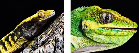
Geckos and anoles are two groups of lizards which possess adhesive toe pads composed of thousands of microscopic hair-like structures (setae) that generate adhesion when placed against a surface. These lizards can be found in a wide variety of habitats and move about on numerous surfaces that differ in roughness, softness, and chemistry. Our labs are interested in understanding how these lizards adhere to biologically-relevant surfaces, such that we can apply this information to the design of synthetic adhesives that can stick under a wide array of conditions. Here are some potential projects in development:
How does setal shape vary across anoles?
In this project, the setae of various anole species will be viewed under a scanning electron microscope and physical measurements taken to better understand the variation in setal shape and configuration.
How does the adhesion of geckos differ on surfaces of varying roughness?
Here, the adhesion of live geckos will be measured on surfaces with different degrees of roughness to obtain data on how roughness affects lizard adhesion.
How does adhesive ability impact the behavior of geckos?
In this project, we will measure gecko adhesion on a variety of rough surfaces and observe
gecko behavior to determine if adhesive ability is related to the types of surfaces geckos move on.
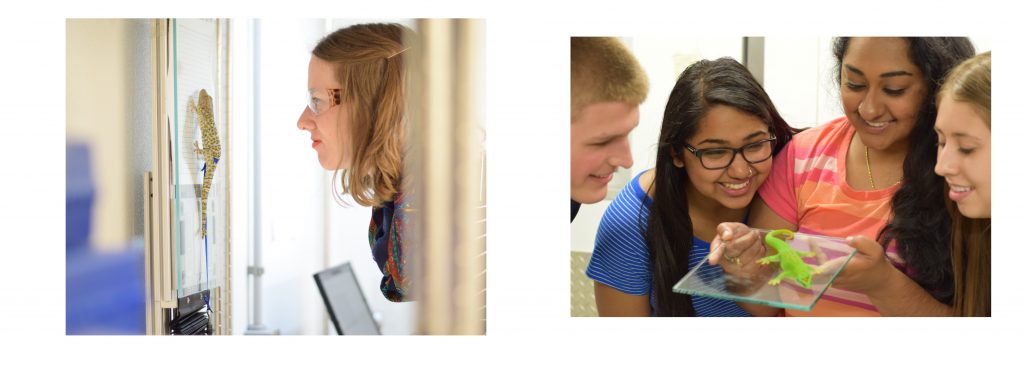
Benefits of this Research Experience
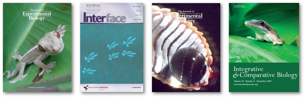
Undergraduate students in the Niewiarowski and Dhinojwala labs will gain an array of critical research skills including: reptile care and handling, live animal performance measurements, experimental design, statistics, microscopy, use of museum collections, surface characterization methods, scientific writing, and more. Additionally, many of the undergraduate students in our labs have been co-authors on several papers published in peer-reviewed journals (see below)!
*Denotes undergraduate student
Niewiarowski, P. H., Lopez, S., Ge, L., Hagan, E.* and Dhinojwala, A. (2008). Sticky gecko feet: the role of temperature and humidity. PLoS. ONE3, e2192.
Niewiarowski, P. H., Stark, A., McClung, B.*, Chambers, B.* and Sullivan, T.* (2012). Faster but Not Stickier: Invasive House Geckos Can Out-Sprint Resident Mournful Geckos in Moorea, French Polynesia. J. Herpetol.46, 194-197.
Stark, A. Y., Sullivan, T. W.* and Niewiarowski, P. H. (2012). The effect of surface water and wetting on gecko adhesion. J. Exp. Biol.215, 3080-6.
Stark, A. Y., Badge, I., Wucinich, N. A.*, Sullivan, T. W.*, Niewiarowski, P. H. and Dhinojwala, A. (2013). Surface wettability plays a significant role in gecko adhesion underwater. Proc Natl Acad Sci U S A110, 6340-5.
Stark, A. Y., Wucinich, N. A.*, Paoloni, E. L.*, Niewiarowski, P. H. and Dhinojwala, A. (2014). Self-drying: a gecko’s innate ability to remove water from wet toe pads. PLoS ONE9, e101885.
Stark, A. Y., McClung, B.*, Niewiarowski, P. H. and Dhinojwala, A. (2014). Reduction of water surface tension significantly impacts gecko adhesion underwater. Integr. Comp. Biol54, 1026-33.
Badge, I., Stark, A. Y., Paoloni, E. L.*, Niewiarowski, P. H. and Dhinojwala, A. (2014). The role of surface chemistry in adhesion and wetting of gecko toe pads. Scientific Reports4, 6643.
Stark, A. Y., Ohlemacher, J.*, Knight, A.* and Niewiarowski, P. H. (2015). Run don’t walk: locomotor performance of geckos on wet substrates. J. Exp. Biol.218, 2435-41.
Stark, A. Y., Palecek, A. M.*, Argenbright, C. W., Bernard, C.*, Brennan, A. B., Niewiarowski, P. H. and Dhinojwala, A. (2015). Gecko adhesion on wet and dry patterned substrates. PLoS. ONE10, e0145756.
Stark, A. Y., Dryden, D. M., Olderman, J.*, Peterson, K. A., Niewiarowski, P. H., French, R. H. and Dhinojwala, A. (2015). Adhesive interactions of geckos with wet and dry fluoropolymer substrates. J. R. Soc. Interface12, 20150464.
Stark, A. Y., Subarajan, S.*, Jain, D., Niewiarowski, P. H. and Dhinojwala, A. (2016). Superhydrophobicity of the gecko toe pad: biological optimization versus laboratory maximization. Phil. Trans. R. Soc. A374, 20160184.
Klittich, M. R., Wilson, M. C., Bernard, C.*, Rodrigo, R. M.*, Keith, A. J.*, Niewiarowski, P. H. and Dhinojwala, A. (2017). Influence of substrate modulus on gecko adhesion. Scientific Reports7, 43647.Garner, A. M.*, Stark, A. Y., Thomas, S. A. and Niewiarowski, P. H. (2017). Geckos go the Distance: Water’s Effect on the Speed of Adhesive Locomotion in Geckos. J. Herpetol.51, 240-244.
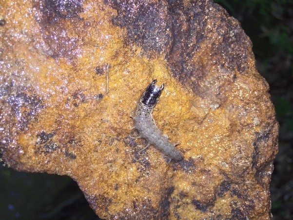
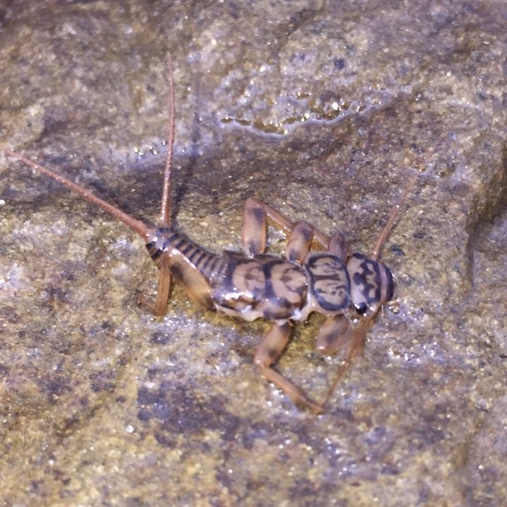
Explore the underwater life in Ohio’s streams, while earning valuable stream assessment skills!
Stream restoration is a growing practice, as people have understood the important ecosystem services that streams provide us. Certain aquatic macroinvertebrates serve as “biological indicators” meaning that their presence or absence indicates the quality of a stream. This research project will be comparing the aquatic macroinvertebrate communities between restored and non- restored streams. As a tiered mentor student, you would assist in the collection of the aquatic macroinvertebrates and learn the Ohio EPA’s methodology for doing so. We will also be surveying stream’s hydrology (flow) and geomorphology (physical channel features). There will also be opportunities to learn aquatic macroinvertebrate identification, data analysis and see some of the area’s beautiful metro parks!
Click here for more information on the Weeks lab
Electronic cigarettes have been in use for over a decade and there is little known about their potential health implications, particularly on embryonic development. We are looking to understand the impact vaping has on cardiovascular development using zebrafish embryos as a model for human embryonic vapor exposure. Zebrafish are a common model organism used in vertebrate developmental research. They are easily maintained, have short developmental time frames, and typically produce large numbers of offspring from a single breeding event. In addition, zebrafish offer the ability to view the developing heart noninvasively, via microscope, due to their lack of body pigmentation during early development. Videos of the beating heart and vasculature can then be recorded and cardiovascular measurements obtained. We are currently seeking one undergraduate student to assist with this project as part of the Tiered Mentoring Program in Biology.
Click here for more information on Dr. Bagatto’s lab.